
Adolescence is a critical developmental phase marked by profound social reorientation, wherein the focus of social engagement shifts dramatically—from the insular family unit to expansive peer networks and institutional environments such as schools. This transition is not merely a social phenomenon but is deeply intertwined with the maturation of the brain’s architecture and functional connectivity. However, for some adolescents, this period presents a paradox: while social engagement is essential, many exhibit behaviors characterized by withdrawal and an increased preference for solitude. Such tendencies are not benign; emerging evidence now suggests they correlate with discernible alterations in brain structure and function, potentially encoding vulnerability to mental health challenges.
In a groundbreaking study led by Dr. Caterina Stamoulis and her team at Boston Children’s Hospital, investigators leveraged the unparalleled depth and breadth of the National Institutes of Health-funded Adolescent Brain Cognitive Development (ABCD) study to unravel the neural substrates underpinning social withdrawal. The ABCD study, encompassing over 11,800 youth across multiple sites in the United States, offers a unique confluence of detailed neuroimaging, behavioral assessments, and environmental profiling. By isolating nearly 3,000 adolescents whose parents provided comprehensive insights into their social behaviors—specifically their proclivity for social withdrawal or solitary preferences—this team was able to pinpoint brain correlates of these behavioral phenotypes with exquisite precision.
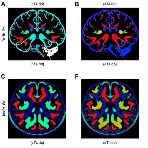
Neuroimaging techniques, including both structural magnetic resonance imaging (MRI) and functional MRI (fMRI), afforded the researchers a dual perspective on the adolescent brain: one that probes anatomical features such as grey matter density and cortical thickness, and another that reveals the oscillatory interplay among discrete neural circuits during rest or task engagement. Adolescents displaying heightened social withdrawal exhibited striking structural deviations primarily localized in brain regions fundamental to social cognition and emotional regulation, notably the insula and anterior cingulate cortex. These areas orchestrate the integration of internal states with external social cues, suggesting that alterations here could underlie the observed behavioral patterns.
Crucially, the deviations extended beyond isolated regions, permeating widespread neural networks. Functional connectivity analyses illuminated diminished coupling strength and increased susceptibility to destabilization within circuits subserving executive functions, decision-making, and affective processing. This widespread neural fragility signals a cascading effect whereby social withdrawal may erode the integrative capacities of the adolescent brain, compromising not only social faculties but also broader cognitive domains. Such diffuse impact provides a compelling mechanistic framework for understanding the heightened risk of mental health disorders linked to persistent social isolation.
Dr. Stamoulis emphasizes that these findings, while confirming theoretical expectations, expose the nuanced reality that solitary behavior in youth is far from a simple byproduct of temperament or choice. Rather, it enacts a profound ripple effect across the neural landscape. The involvement of multiple networks points towards a systemic neurodevelopmental vulnerability, wherein early patterns of social disengagement could precipitate or exacerbate psychopathology. This insight provides an urgently needed biological substrate for what clinicians often observe but cannot easily quantify: the insidious toll of social isolation during a critical period of neuroplasticity.
For pediatricians, psychiatrists, and allied clinicians, the implications are clear and multifaceted. Solitude, in measured doses, is a normative and integral feature of adolescent development, fostering self-reflection and autonomy. Yet when solitude patterns escalate and persist, coinciding with neural alterations, they signal a red flag demanding intervention. Educating families about the neurobiological correlates of social withdrawal transforms anecdotal concerns into tangible risks, fostering proactive engagement. By framing withdrawal within a neuroscientific context, healthcare providers can dismantle stigma, encourage empathy, and galvanize early, tailored support aimed at forestalling downstream mental health sequelae.
The power of the ABCD study resides not only in its scale but also in its longitudinal design. Repeated neuroimaging and behavioral assessments every two years allow researchers to delineate the temporal dynamics of brain development vis-à-vis social behaviors. Dr. Stamoulis and colleagues anticipate that future analyses will illuminate trajectories of risk and resilience, charting how enduring patterns of solitude influence brain maturation or, conversely, how timely interventions recalibrate neural networks in favor of healthier social integration.
Another layer of complexity pertains to disentangling causality from correlation. Does social withdrawal instigate neural changes, or do preexisting brain patterns predispose some adolescents to seek solitude? The longitudinal approach adopted will be instrumental in elucidating these bidirectional influences. Moreover, integrating environmental, psychological, and genetic data stands to enrich this multidimensional portrait, offering precision targets for intervention and prevention.
This research underscores an often-overlooked dimension of adolescent health: the intersection of social environment, brain development, and mental wellness. By leveraging cutting-edge neuroimaging techniques and robust behavioral phenotyping within a large, diverse sample, this study elevates our understanding of solitude from a social curiosity to a measurable brain phenomenon with far-reaching implications. The translational potential is immense—empowering clinicians, educators, caregivers, and policymakers to recognize and address solitude not merely as a behavioral eccentricity but as a sentinel of neurodevelopmental health deserving focused attention and resources.
The study, published in the prestigious journal Cerebral Cortex, marks a pivotal contribution to developmental neuroscience and psychiatry. It highlights the exigency of multidimensional research modalities that encompass not only observable behaviors but also their covert neural signatures. Moreover, by drawing upon the resources of the National Science Foundation’s Developmental Sciences and Collaborative Research in Computational Neuroscience programs, this work exemplifies the synergy between large-scale funding and transformative scientific discovery.
In summary, the journey from adolescence into adulthood is punctuated by a delicate balance of social engagement and solitary reflection. When the scales tip toward withdrawal, the adolescent brain echoes this shift with structural and functional repercussions that transcend mere social discomfort. Dr. Stamoulis’s team has charted these neural alterations, laying the groundwork for future endeavors aimed at mitigating the shadow cast by social isolation. As the ABCD study progresses, the promise of early detection and intervention shines brighter, heralding a new era where solitary behaviors are understood, monitored, and managed with the sophistication they warrant.
Subject of Research: Neural correlates of social withdrawal and preference for solitude during adolescence.
Article Title: Neural Correlates of Social Withdrawal and Preference for Solitude in Adolescence.
News Publication Date: October 2, 2025.
Web References: https://abcdstudy.org/; http://dx.doi.org/10.1093/cercor/bhaf260
References: Published article in Cerebral Cortex, DOI: 10.1093/cercor/bhaf260.
Keywords: Adolescents, Social withdrawal, Cognitive development, Cognitive function, Neuroimaging, Psychological science.

