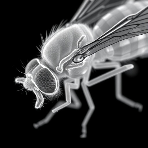
In an extraordinary leap forward for our understanding of cellular filtration systems, a recent study published in Nature Communications reveals the intricate architecture of the slit diaphragm in Drosophila, the common fruit fly. This specialized cellular structure, long known to be integral in filtration processes within the kidney-like nephrocytes of insects, has now been visualized in unprecedented detail. The researchers illuminate a bilayered, fishnet-like configuration, challenging and expanding existing paradigms about how molecular sieving is orchestrated at the nanoscale level within these remarkably thin filters.
The slit diaphragm has fascinated biologists for decades due its pivotal role in selectively filtering blood plasma, maintaining homeostasis, and preventing the leakage of vital proteins. Yet until now, understanding of its precise structural organization remained elusive. Utilizing state-of-the-art imaging techniques, including high-resolution electron tomography and advanced molecular modeling, the authors dissect the spatial arrangement of proteins forming this labyrinthine network. Their findings not only decode the fundamental blueprint of the slit diaphragm but also hint at evolutionary conservation across species, suggesting a universal principle in biological filtration.
At the core of this research lies the discovery that the slit diaphragm is not a single-layered mesh as previously assumed but consists of two distinct layers woven into an interlocking fishnet pattern. This bilayered assembly presumably fortifies the structure against mechanical stress while preserving flexibility to adapt shape in response to hemodynamic forces. Such dual-layer construction could explain how slit diaphragms efficiently maintain selective permeability, allowing essential solutes to pass while blocking larger molecules and potential pathogens, thus safeguarding cellular and systemic health.
The intricate fishnet architecture appears to be formed primarily by the interaction of transmembrane proteins, including orthologs of nephrin and NEPH1, which have been documented in mammalian kidneys. Through meticulous mapping of these molecular constituents and their interface, the study demonstrates how they interlace vertically and laterally to generate a robust yet dynamic barrier. This structural insight bridges gaps in previously disconnected data sets, offering a coherent model that aligns molecular composition with mechanical and functional attributes.
Moreover, the researchers unveil that this bilayer consists of two staggered planes of protein complexes, each contributing distinctive biochemical properties and anchorage points. The upper and lower layers connect through specialized linker domains, ensuring resilience against physical perturbations such as stretch and shear forces induced by blood flow. This mechanical coupling paradigm is analogous to engineered nets used in human technology but optimized by evolution for continuous, highly selective filtration under fluctuating physiological conditions.
This pioneering work carries profound implications for nephrology and beyond. Understanding the slit diaphragm’s refined architecture opens new avenues towards comprehending pathologies like proteinuric kidney diseases, where the filtration barrier is compromised. Mutations in slit diaphragm constituents lead to catastrophic breakdowns, resulting in chronic kidney dysfunction. The Drosophila model, with its conserved slit diaphragm design, therefore provides a powerful platform for dissecting molecular mechanisms underlying these disease states and screening therapeutic interventions.
In parallel, this fishnet-like bilayer could inspire biomimetic material science, where synthetic filtration membranes emulate biological efficiency and specificity. Engineers might replicate this dual planar system to design adaptive filters for medical devices, water purification, or molecular sieves that demand precision and structural durability in a compact form factor. The study’s granular characterization of linker domains and protein interfaces could guide the molecular engineering required to achieve such biomimicry.
The team behind this study harnessed cutting-edge correlative light and electron microscopy to visualize nephrocytes in their native tissue context. This integrative approach preserved physiological architecture while achieving molecular resolution, a feat unattainable by traditional techniques alone. By combining spatial and chemical information, the researchers could pinpoint how specific proteins distribute across the bilayer and interact dynamically, an essential step towards causal mechanistic deciphering.
This work also prompts a reevaluation of the evolutionary trajectory of filtration systems. Insects and vertebrates diverged approximately 500 million years ago, yet their slit diaphragms share architectural motifs and key molecular players. The bilayered, fishnet design likely represents a fundamental evolutionary solution to the universal challenge of molecular filtration under fluid mechanical stress. This conservation underscores how natural selection can shape and iterate on highly efficient nanoscale structures across vast phylogenetic distances.
Furthermore, the comprehensive structural model allowed the authors to simulate mechanical responses under various conditions mimicking physiological forces. These biophysical simulations revealed how the bilayer arrangement dissipates and distributes mechanical loads, mitigating risks of tears or ruptures. Such resilience is crucial for long-term function in an organ constantly exposed to pressure fluctuations, impinging flow, and biochemical insults. The insights gleaned here add a new dimension to understanding slit diaphragm homeostasis and repair mechanisms.
In addition to advancing fundamental biology, this study revitalizes interest in Drosophila as a model organism for kidney function research. The genetic tractability of flies combined with high-resolution structural insights positions Drosophila nephrocytes to become a central system for investigating nephrotic syndromes, drug-induced nephrotoxicity, and regenerative biology. This fusion of genetics, imaging, and biophysics exemplifies a multidisciplinary approach that will reshape renal science.
Notably, the detailed characterization of the slit diaphragm’s adhesive properties highlights how specific protein domains mediate tight yet reversible binding. This fine-tuned balance facilitates dynamic remodeling during development and stress while preserving filter integrity. Such regulatory mechanisms likely contribute to the slit diaphragm’s ability to self-heal microscopic damages, preventing cumulative filtration failure—a remarkable feat of biological engineering with tantalizing parallels in tissue engineering.
From a therapeutic perspective, the elucidation of bilayer formation mechanisms points to potential molecular targets. Modulating interactions between nephrin, NEPH1, and linker proteins could restore barrier function in diseased states or prevent damage progression. The structural framework provided by this study paves the way for rational drug design, guiding precision interventions tailored to specific molecular defects underlying proteinuria and nephrotic diseases.
Moreover, by mapping the fishnet arrangement in exquisite detail, the authors provide a template for reconstructing functional slit diaphragms in vitro. This capability could transform drug testing and disease modeling by offering platforms that recapitulate physiological filtration barriers faithfully. Such ex vivo systems could accelerate discovery and reduce reliance on animal testing, marking a significant advance in biomedical research infrastructure.
The implications of this research ripple beyond nephrology, as slit diaphragm-like structures appear in diverse organisms fulfilling selective barrier functions. Decoding the principles of bilayer fishnet design elucidates nature’s broader strategies for balancing selectivity, permeability, and mechanical robustness. These lessons inform disciplines from developmental biology to bioengineering, illustrating how nanoscale architecture governs emergent biological function with remarkable efficiency.
Ultimately, this landmark study presents the slit diaphragm as a masterpiece of biological design—a sophisticated net woven by evolution into a seamless, bilayered filter. Its revelation reshapes our conceptions of cellular filtration and opens thrilling new frontiers across medicine, biology, and material science. As researchers continue to unravel its secrets, the humble fruit fly delivers profound insights that may transform human health and technology in ways we are only beginning to imagine.
Subject of Research: The structural organization and molecular architecture of the slit diaphragm in Drosophila nephrocytes.
Article Title: The slit diaphragm in Drosophila exhibits a bilayered, fishnet architecture.
Article References:
Moser, D., Lang, K., Birtasu, A.N. et al. The slit diaphragm in Drosophila exhibits a bilayered, fishnet architecture. Nat Commun 16, 8741 (2025). https://doi.org/10.1038/s41467-025-64347-5
Image Credits: AI Generated
Tags: advanced molecular modeling techniquesbilayered fishnet architecturebiological filtration principlescellular filtration systemsDrosophila slit diaphragm structureevolutionary conservation in filtration systemshigh-resolution electron tomographykidney-like structures in insectsmolecular sieving at nanoscalenephrocytes in insectsprotein spatial arrangement in filtersselective blood plasma filtration


