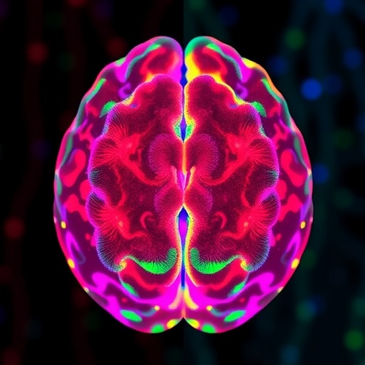
In a transformative leap for neuroscience and medical imaging, researchers have unveiled a pioneering technique that enables simultaneous, large-scale visualization of neuronal, astrocytic, and hemodynamic activities within the living brain. This hybrid imaging modality, termed Hybrid Multiplexed Fluorescence and Magnetic Resonance Imaging (HyFMRI), represents a paradigm shift in non-invasive brain research, offering unprecedented insight into the complex interplay between diverse cellular and vascular processes in real time.
At the heart of this innovation lies the integration of multiplexed fluorescence imaging, which can distinguish the activities of neurons and astrocytes by tagging these cells with distinct fluorescent markers, with the comprehensive spatial resolution of magnetic resonance imaging (MRI). By fusing these complementary imaging techniques, HyFMRI allows researchers to simultaneously capture biochemical and physiological dynamics across wide brain regions without the limitations imposed by traditional methods that usually focus on isolated elements or require invasive procedures.
The novel approach addresses a critical gap in neuroimaging: capturing concurrent functional signals from multiple cell types while monitoring their hemodynamic context. Understanding these dynamics is essential because neurons rely not only on electrical impulses but also on astrocytic support and vascular responses to sustain complex brain functions. Previous imaging techniques have struggled to provide a holistic view, often focusing exclusively on either neuronal activity or blood oxygenation level-dependent (BOLD) signals, leaving astrocytes—and their role in neurometabolic coupling—largely elusive.
HyFMRI leverages advanced fluorescent reporter proteins engineered to respond to electrical and calcium signals specifically in neurons and astrocytes. These reporters enable differentiation and tracking of cellular activities in vivo. Meanwhile, the MRI component delivers volumetric data on blood flow and oxygenation, bridging a critical link between cellular signaling and vascular responses. The simultaneous acquisition of these datasets facilitates the mapping of neurovascular coupling with high temporal and spatial fidelity.
One of the standout capabilities of HyFMRI is its non-invasive application, which crucially preserves the integrity of the brain’s microenvironment. Unlike invasive electrophysiological methods or fluorescence microscopy restricted to superficial layers, this technique probes deeper structures while maintaining broad coverage. This attribute is especially valuable for longitudinal studies monitoring disease progression, therapeutic responses, or neurodevelopmental processes over extended periods.
The technical synergy was achieved by designing a specialized imaging setup synchronized to coordinate the excitation and emission of multiplexed fluorescent signals alongside MRI data acquisition sequences. This coordination mitigates signal cross-talk and artifact formation that could otherwise degrade image quality. Moreover, innovative computational algorithms process and integrate the multimodal data in real time, enhancing signal extraction and enabling dynamic correlation analyses of neural, astrocytic, and vascular interactions.
Preclinical applications in rodent models demonstrated the method’s prowess. The team was able to visualize stimulus-evoked neuronal firing patterns concurrently with astrocytic calcium waves and corresponding hemodynamic fluctuations. These findings underscore the interdependence of cellular and vascular responses, furnishing critical clues to underlying mechanisms in sensory processing and brain energetics, thereby advancing our understanding of fundamental brain function.
Importantly, HyFMRI holds the promise to revolutionize the study of neurological disorders where aberrant neurovascular coupling and astrocyte dysfunction have been implicated, including Alzheimer’s disease, stroke, epilepsy, and neuroinflammation. By providing detailed spatiotemporal maps of pathological alterations in cellular and vascular dynamics, this method offers a powerful tool for early diagnosis, monitoring, and the evaluation of therapeutic interventions.
Beyond clinical implications, the ability to visualize simultaneous activities of neurons and astrocytes alongside cerebral hemodynamics offers a richer canvas for neuroscience research. It can illuminate the roles astrocytes play in modulating synaptic activity, plasticity, and neuronal metabolism within intact networks. This could reshape prevailing models that historically marginalized glial cells to mere support roles, highlighting their active participation in brain computations.
The researchers also emphasize the technique’s adaptability. HyFMRI could be tailored to target various cellular markers beyond neurons and astrocytes by incorporating additional fluorescent probes. Such flexibility extends its applications to diverse studies involving microglia, oligodendrocytes, or even genetically encoded biosensors reporting neurotransmitters or metabolic states, thus expanding its utility across neuroscience disciplines.
While the current iteration mainly targets rodent models, efforts are underway to refine HyFMRI for potential human applications. Challenges including scaling the fluorescence detection sensitivity and adapting MRI protocols for clinical scanners are active areas of development. The eventual translation of this technology to human neuroimaging could transform diagnostics and research, enabling non-invasive, multi-modal monitoring of brain health and disease with cellular resolution.
This breakthrough also stimulates the dialogue surrounding multimodal imaging integration. The successful marriage of fluorescence multiplexing with MRI offers a blueprint for future innovations combining optical and magnetic resonance technologies, encouraging the exploration of new hybrid systems. Such interdisciplinary advancements rely on collaboration across bioengineering, optics, neurobiology, and medical imaging fields.
Ultimately, HyFMRI exemplifies the power of convergent technologies to disentangle the brain’s complexity. By illuminating the concurrent dynamics of neuronal activity, astrocytic signaling, and vascular responses, scientists now possess a more holistic lens to decode brain function. This advancement brings us closer to comprehending how cellular interplay orchestrates cognition, behavior, and neuropathology in the living brain.
The study, published in Light: Science & Applications, marks a milestone in neuroimaging that could redefine brain research in the years to come. It extends beyond mere imaging innovation, offering a versatile platform poised to accelerate discoveries in neuroscience and medicine. As further refinements and applications emerge, HyFMRI may soon become indispensable in laboratories and clinics worldwide.
Intriguingly, the hybrid system provides rich, multidimensional datasets that also invite the integration of artificial intelligence and machine learning algorithms. These tools can dissect the complex spatiotemporal patterns uncovered by HyFMRI, facilitating automated identification of network states, prediction of disease trajectories, or personalized therapeutic adjustments, pushing the frontiers of precision neuroscience.
In conclusion, Hybrid Multiplexed Fluorescence and Magnetic Resonance Imaging sets a new standard for functional brain imaging. Its capacity to concurrently capture multi-cellular signaling alongside vascular dynamics non-invasively heralds a transformative era in brain research. This work underscores the potential of hybrid imaging modalities to unravel the brain’s inner workings with unprecedented clarity and scale.
Subject of Research: Hybrid neuroimaging techniques integrating multiplexed fluorescence and magnetic resonance imaging for simultaneous detection of neuronal, astrocytic, and hemodynamic activity.
Article Title: Non-invasive large-scale imaging of concurrent neuronal, astrocytic, and hemodynamic activity with hybrid multiplexed fluorescence and magnetic resonance imaging (HyFMRI).
Article References:
Chen, Z., Chen, Y., Gezginer, I. et al. Non-invasive large-scale imaging of concurrent neuronal, astrocytic, and hemodynamic activity with hybrid multiplexed fluorescence and magnetic resonance imaging (HyFMRI). Light Sci Appl 14, 341 (2025). https://doi.org/10.1038/s41377-025-02003-9
Image Credits: AI Generated
DOI: https://doi.org/10.1038/s41377-025-02003-9
Tags: brain activity visualizationcellular dynamics in neurosciencehemodynamic activity monitoringhybrid imaging techniquesHyFMRI technologyinterdisciplinary neuroscience researchmagnetic resonance imaging applicationsmultiplexed fluorescence imagingneuroimaging advancementsneuronal astrocytic interactionsnon-invasive brain researchreal-time brain activity analysis


