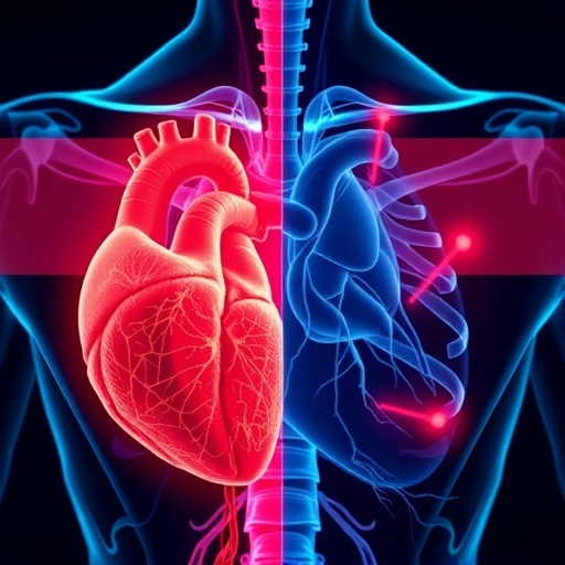
In a groundbreaking study that reshapes our understanding of cardiovascular neurobiology, researchers at the University of Bristol have unveiled striking asymmetrical adaptations in the nerves controlling the heart in response to aerobic exercise. This new research, published in the prestigious journal Autonomic Neuroscience, reveals for the first time that moderate aerobic training induces a side-specific remodeling of the stellate ganglia—the paired nerve clusters that exert profound influence over cardiac function. These findings could herald a new era in how we approach treatment for a spectrum of heart conditions, from arrhythmias to angina and stress-related cardiomyopathies.
Exercise is widely recognized as a cornerstone of heart health, well-known for strengthening cardiac muscle and improving vascular function. However, the University of Bristol-led team has now revealed a deeper, more nuanced impact: moderate aerobic activity causes the autonomic nervous system—often dubbed the body’s “autopilot”—to adjust its control architecture in a lateralized manner. Their investigation details how the structural neuroplasticity in the stellate ganglia differs markedly between the left and right sides, suggesting a complex neuro-regulatory system responsive to lifestyle factors.
The stellate ganglia are small but critically important nerve hubs located in the lower neck and upper chest region that dispatch sympathetic signals orchestrating heart rate and contractility. By leveraging advanced three-dimensional quantitative imaging techniques known as stereology, the research team quantified changes in neuronal populations in these ganglia in rat models subjected to a 10-week aerobic training regimen. The results were striking: the right stellate ganglion exhibited a remarkable increase in neuron number—about four times greater than in sedentary controls—while the left ganglion neurons did not proliferate but instead underwent hypertrophy, nearly doubling in size.
This asymmetric neuroplasticity is of particular significance considering the distinct functional roles historically attributed to the left and right stellate ganglia. Prior clinical observations hinted at laterality influencing cardiac pathophysiology and treatment efficacy, but the underlying anatomical and physiological bases were elusive. These newly documented side-specific structural changes may therefore provide an anatomical explanation for why certain cardiac interventions, such as nerve blocks or targeted denervations, yield disparate outcomes depending on the side treated.
Dr. Augusto Coppi, Senior Lecturer in Veterinary Anatomy at the University of Bristol and the study’s lead author, emphasized the importance of these findings: “Our research exposes a previously hidden pattern in the heart’s autonomic regulation system. The left-right divergence in neural remodeling suggests that exercise doesn’t merely strengthen the heart—it rewires its neural control with precision.” He further postulated that these lateralized adaptations might inform personalized therapeutic strategies, optimizing interventions for conditions such as arrhythmias and angina by targeting the side most likely to yield benefit.
Beyond the structural adaptations, the implications stretch into pathophysiological domains. Many prevalent cardiac disorders stem from dysregulation of sympathetic nerve activity mediated through the stellate ganglia. For example, stress-induced cardiomyopathy—commonly known as ‘broken-heart syndrome’—and certain refractory arrhythmias are associated with overactive sympathetic signaling. Understanding how aerobic exercise reshapes these sympathetic nerve clusters holds the promise of fine-tuning medical procedures that aim to modulate nerve activity, potentially improving patient outcomes while reducing invasive intervention frequency.
This study also opens avenues for translational research exploring how these side-specific modifications in autonomic nerve architecture map onto functional cardiac outcomes in humans. The research team plans to conduct follow-up studies tracing correlations between these neuroplastic changes and measurable differences in heart rhythm and contractile behavior at rest and during exertion. Preliminary data in rats provide a compelling rationale to investigate these mechanisms in larger animal models and human subjects, utilizing advanced non-invasive imaging and electrophysiological monitoring technologies.
The collaborative project involved expertise from University College London as well as University of São Paulo and Federal University of São Paulo in Brazil, demonstrating the interdisciplinary and international nature of cutting-edge cardiovascular neuroscience. The utilization of stereology for 3D neuronal mapping underscores the precision required to discern subtle yet impactful neuroanatomical changes, paving the way for future research into the autonomic nervous system’s plasticity across different organ systems.
Intriguingly, the left stellate ganglion’s neuronal hypertrophy contrasted with the right’s increase in neuron numbers suggests divergent cellular mechanisms underlying the plastic response. Hypertrophy, involving enlargement of existing neurons, may correspond to enhanced synaptic strength or functional efficacy, whereas increased neuron counts on the right side imply neurogenesis or neural proliferation. Unpacking these cellular phenomena will be a pivotal next step in understanding how exercise influences the nervous regulation of the heart.
The identification of lateralized neural remodeling also aligns with a growing body of research emphasizing the brain and autonomic nervous system’s lateralization in controlling physiological processes. While such neurological asymmetry is well documented in higher cognitive functions, its manifestation in peripheral autonomic ganglia controlling cardiac function offers novel insights into the complexity of neurocardiac interplay.
As modern medicine moves towards individualized treatments based on patient-specific anatomical and functional profiles, the discovery of side-specific autonomic nerve remodeling represents a significant advance. Future clinical trials designed with this asymmetry in mind could transform approaches to interventions like stellate ganglion blocks and selective denervation, potentially tailoring treatments to maximize efficacy and minimize side effects.
Moreover, the findings may hold implications beyond cardiovascular disease. Since sympathetic nervous system overactivity is implicated in various systemic disorders, from hypertension to heart failure, understanding how lifestyle factors such as exercise induce neuroplasticity could inspire holistic therapeutic approaches extending beyond standard pharmacology.
With this research, Dr. Coppi and colleagues have laid essential groundwork illuminating how regular moderate aerobic exercise remodels the nervous system controlling the heart in a side-specific manner. Their work not only deepens scientific comprehension of heart-nerve interactions but also challenges clinicians and researchers to rethink treatment paradigms, embracing neuroanatomical asymmetry as a critical factor in cardiovascular health and disease management.
Subject of Research: Animals
Article Title: ‘Asymmetric neuroplasticity in stellate ganglia: unveiling side-specific adaptations to aerobic exercise’
News Publication Date: 23-Sep-2025
Web References: 10.1016/j.autneu.2025.103338
Keywords: Human health
Tags: aerobic exercise and heart healthasymmetrical adaptations in heart nervesautonomic nervous system and exercisecardiovascular neurobiology researchheart control nerves remodelinglifestyle factors affecting heart healthmoderate aerobic training effects on heartneuroplasticity in stellate gangliaregular exercise benefitsside-specific cardiac function adaptationstreatment implications for heart conditionsUniversity of Bristol cardiovascular study


