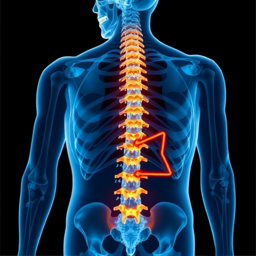
In a groundbreaking study published in the Journal of Medical Biology and Engineering, researchers have extensively analyzed the biomechanical effects of defect size and location on vertebral fractures. This research emerges from a pressing need to understand how different variables impact vertebral integrity, a topic of significant concern in the fields of orthopedics and sports medicine. As humanity continues to live longer, the prevalence of vertebral fractures, particularly in the elderly population, has prompted renewed investigation into the biomechanics of the spinal architecture.
The vertebral column, a complex structure composed of vertebrae, intervertebral discs, ligaments, and muscles, bears the weight of the upper body while facilitating movement. Each vertebra is designed to absorb and distribute loads effectively. However, when defects arise due to conditions such as osteoporosis or traumatic injury, the biomechanical characteristics can be significantly altered, leading to an increased risk of fractures. This latest work by Rezaei and colleagues aims to demystify the relationship between defect size, location, and the resulting biomechanical responses.
The methodology employed in this study was robust, utilizing a combination of computational modeling and experimental validation. Advanced finite element analysis (FEA) techniques were applied to simulate various defect scenarios in vertebrae. This allowed researchers to quantitatively assess the stress distribution patterns and failure modes associated with different defect configurations. These simulations provided critical insights into how specific defect sizes and their locations on the vertebra influence its mechanical performance.
One of the pivotal findings was that defect size plays a crucial role in determining the fracture risk of vertebrae. The researchers found that larger defects significantly compromised the mechanical integrity of the vertebra, leading to increased localized stress concentrations. This finding is particularly important for clinicians who assess patient risks for spinal fractures and can lead to better personalized treatment strategies. The data suggest that conservative management may be more appropriate for smaller defects, while larger defects may necessitate surgical intervention.
Additionally, the study unveiled intriguing insights regarding the location of defects. Defects located in posterior regions of the vertebral body presented a greater risk of fractures compared to anterior defects. This information underscores the importance of understanding the anatomical context of lesions and how their positioning impacts biomechanical stability. It also raises questions about the mechanical behavior of the vertebrae under different loading conditions, which may be relevant in sports medicine or trauma care.
The clinical implications of this research extend beyond academic interest. By informing clinicians about the biomechanical risks associated with different types of vertebral defects, it may enhance decision-making processes in surgical planning and rehabilitation protocols. The researchers also suggest that monitoring the size and location of vertebral defects could lead to more effective preventive measures in vulnerable populations.
Moreover, the integration of these findings into patient assessments can refine the understanding of osteoporosis management, a condition that significantly affects the elderly. As osteoporosis can lead to both the formation of defects and increased fracture risk, this study provides a valuable framework for holistic patient management. Preventative strategies that consider both defect characteristics and general bone health can be more effectively developed.
The research team also acknowledged the limitations of their study, suggesting that while the FEA results are robust, clinical applications will require further validation in in vivo settings. Future research may include in real-time biomechanics during various activities that reflect daily living or athletic performances, wherein the effects of dynamic loading can be studied.
In related advancements, the use of imaging techniques, such as MRI or CT scans, could significantly improve the detection and characterization of vertebral defects. By incorporating imaging data with biomechanical simulations, practitioners may gain a comprehensive view of a patient’s spinal health and fracture risk. The confluence of technology and medicine is paving the way for innovative diagnostic and treatment methodologies.
It is the fusion of biological understanding and engineering principles that defines the cutting edge of orthopedic research. The interplay between size, location, and mechanical properties of spinal defects will advance our grasp of vertebral health and injury prevention. As this research gains traction, awareness of its significance in clinical practices will undoubtedly increase.
The implications of this study span multiple disciplines. From biomechanics to medical imaging, the collaborative efforts across fields will continue to shape the future of patient care and treatment modalities for vertebral fractures. With further validation and exploration of these findings, one can anticipate novel interventions that could drastically reduce the incidence and consequences of vertebral fractures, ultimately improving patient quality of life.
Researchers emphasize the need for more comprehensive studies that could include demographic variations, different loading scenarios, and the potential impact of rehabilitation techniques on the healing of vertebral defects. As the scientific community grapples with these complex issues, the importance of multidisciplinary approaches becomes even clearer.
In conclusion, the research conducted by Rezaei et al. sheds light on a critical issue in spinal health. By systematically investigating the biomechanical effects of defect size and location, the findings represent a significant advancement in our understanding of vertebral fractures. This study lays the groundwork for further research that may lead to more nuanced guidelines for clinical practice, emphasizing the need for ongoing innovation in spinal health management.
Subject of Research: Biomechanical effects of defect size and location on vertebral fractures.
Article Title: Biomechanical Effects of Defect Size and Location on Vertebral Fracture.
Article References:
Rezaei, A., Schreiber, A., Dashtdar, B. et al. Biomechanical Effects of Defect Size and Location on Vertebral Fracture.
J. Med. Biol. Eng. (2025). https://doi.org/10.1007/s40846-025-00976-x
Image Credits: AI Generated
DOI: 10.1007/s40846-025-00976-x
Keywords: vertebral fracture, biomechanics, finite element analysis, defect size, defect location, osteoporosis, spinal health.
Tags: biomechanical responses to spinal defectsbiomechanics of spinal architecturecomputational modeling of vertebral defectsdefect size impact on spinal fracturesexperimental validation in biomechanics researchfinite element analysis in orthopedicslocation effects on vertebral integrityorthopedic implications of vertebral fracturesosteoporosis and spinal injuriessports medicine and spinal healthvertebral column load distributionvertebral fractures in elderly population

