In a groundbreaking pilot study, researchers have illuminated the potential of ultra-low-dose computed tomography (ULDCT) as an innovative imaging modality to unveil the often elusive structural airway abnormalities in adolescents and young adults living with cystic fibrosis (CF). This progressive condition, characterized by persistent lung infections and declining pulmonary function, demands precise and minimally invasive diagnostic tools to tailor and monitor treatment efficacy. The novel application of ULDCT, combined with patient-centered co-production approaches, marks a significant leap forward in both technological advancement and therapeutic engagement for people with cystic fibrosis (PwCF).
Cystic fibrosis is a complex genetic disorder that primarily affects the respiratory and digestive systems, with pulmonary disease as the leading cause of morbidity and mortality. The management of CF has evolved dramatically with the introduction of highly effective modulators such as elexacaftor/tezacaftor/ivacaftor (ETI), which target the underlying CF transmembrane conductance regulator (CFTR) protein dysfunction. Despite these pharmacological breakthroughs, monitoring structural airway damage remains essential to optimize airway clearance therapy (ACT) and identify disease progression or treatment response. Herein lies the transformative promise of ULDCT—a technique that drastically reduces radiation exposure compared to conventional CT scans, making serial imaging safer and more feasible for younger populations.
The pilot study undertaken by Zhang et al. investigates not only the technical feasibility of employing ULDCT in detecting intricate airway changes but also explores the psychosocial impact of integrating imaging results with patients’ involvement in their care. This multifaceted approach leverages co-production, a collaborative process where PwCF actively participate in interpreting their imaging findings alongside clinicians. Such engagement is hypothesized to catalyze improved adherence to ACT regimens and enhance overall patient satisfaction by directly linking visual evidence of airway health to therapeutic actions.
From a technical perspective, ULDCT employs advanced image reconstruction algorithms and optimized scanning protocols to achieve high-resolution images at radiation doses comparable to or lower than a chest x-ray. The precise calibration of parameters such as voltage, current, and acquisition time enables clinicians to capture detailed cross-sectional images of airway structures while mitigating long-term radiation risks, a crucial consideration in lifelong diseases requiring repeated imaging. This delicate balance between diagnostic clarity and patient safety positions ULDCT as a potentially transformative imaging tool in the realm of pediatric and young adult pulmonology.
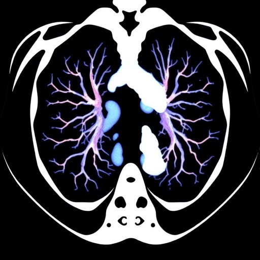
The image displayed in the study reveals the striking clarity with which ULDCT delineates bronchial wall thickening, mucus plugging, and bronchiectasis—hallmarks of CF lung disease—providing invaluable insights into the extent and distribution of airway pathology. These structural abnormalities, often subtle or difficult to discern with less sensitive imaging modalities, are critical markers for disease staging and monitoring. The ability of ULDCT to detect such changes with minimal radiation exposure means that clinicians can now consider more frequent assessments, thereby enabling a more dynamic and responsive treatment strategy.
Beyond the technical triumphs, the study underscores a vital shift towards patient empowerment through co-production. This novel model fosters a more transparent and interactive clinical relationship, wherein patients and providers review ULDCT images together, discuss the implications of detected abnormalities, and collaboratively develop or adjust airway clearance plans. This shared decision-making framework not only demystifies complex radiological data for patients but also embeds personalized care at the heart of CF management.
Initial findings suggest that when PwCF visualize tangible evidence of airway damage, their motivation to adhere to rigorous ACT protocols significantly improves. By bridging the gap between abstract clinical advice and real-world physiological manifestations, ULDCT review sessions elicit a profound behavioral impact, potentially enhancing long-term clinical outcomes. Moreover, patient-reported satisfaction with their treatment routine appears to increase, likely due to heightened understanding and a sense of active involvement in their disease management.
A central advantage of ULDCT lies in its compatibility with the clinical needs of CF patients undergoing ETI therapy. While ETI has revolutionized the treatment landscape by improving lung function and halting disease progression, subtle structural changes can persist or evolve. Traditional imaging modalities may fail to capture these nuances early enough to prompt therapeutic adjustments. ULDCT, therefore, serves as a critical adjunct, furnishing precise anatomical data that complement functional assessments such as spirometry, thereby offering a more comprehensive view of patient status.
The implications of this pilot study extend beyond CF care; they herald a paradigm shift in chronic respiratory disease management where low-risk imaging coupled with patient engagement can redefine therapeutic adherence. As medical practice gravitates towards precision medicine, the integration of cutting-edge diagnostics with collaborative care models epitomizes a future where patients are informed partners rather than passive recipients—engaged, empowered, and equipped to make meaningful decisions about their health.
Researchers acknowledge certain limitations inherent to the pilot nature of their study, including a modest sample size and the need for longitudinal data to validate ULDCT’s prognostic value and reproducibility. However, the promising results pave the way for larger, multicenter trials designed to refine scanning protocols further and explore the psychosocial dynamics of co-production in greater depth. Future research may also investigate the economic impact of widespread ULDCT adoption, weighing the benefits of improved disease monitoring against costs and resource allocation.
Technical challenges addressed in the study include optimizing image quality despite the ultra-low radiation dose and ensuring consistent interpretation across radiologists with varying experience levels. The research team employed state-of-the-art iterative reconstruction techniques and standardized image scoring systems to enhance diagnostic reliability. Furthermore, the study highlights the importance of multidisciplinary collaboration—radiologists, pulmonologists, physiotherapists, and patient advocates all play pivotal roles in interpreting images, fostering dialogue, and adapting treatments.
In addition to structural evaluation, ULDCT may hold potential for assessing airway inflammation and infection patterns when coupled with emerging imaging biomarkers in the future. Such advancements could amplify the modality’s utility, allowing clinicians to visualize not only anatomical but also pathophysiological changes, thereby guiding more tailored interventions. The integration of artificial intelligence in image analysis is another frontier anticipated to enhance the sensitivity and specificity of ULDCT, streamlining workflows and providing real-time decision support.
The study by Zhang and colleagues sets a precedent for embedding patient-centered technologies within routine clinical practice, especially in diseases where treatment complexity and chronic management impose significant burdens. The behavioral insights gained from co-produced imaging review sessions could inform strategies across a spectrum of chronic illnesses, amplifying adherence and satisfaction universally. This approach respects patient autonomy, fosters trust in healthcare teams, and exemplifies a culture of transparent care grounded in evidence.
As the prevalence of CF continues to decline due to therapeutic advancements, the focus transitions toward optimizing quality of life and minimizing cumulative treatment toxicity. The emerging role of ULDCT in this context is emblematic of precision care—noninvasive, patient-friendly, yet comprehensive in capturing the pathological footprint of disease. Its adoption could shorten the diagnostic lag, unmask early airway degradation, and foster proactive clinical decision-making, ultimately altering the disease trajectory and enhancing survivorship.
In conclusion, this innovative pilot study pioneers the intersection of advanced imaging technology and co-production in cystic fibrosis care. By demonstrating the feasibility and clinical promise of ULDCT combined with active patient engagement, Zhang et al. chart a path toward more nuanced, personalized, and patient-centric respiratory care. As the field embraces this dynamic, the ripple effects are poised to transform not only CF management but broader paradigms of chronic illness treatment, underscoring the power of merging technical innovation with human collaboration.
Subject of Research: The feasibility and impact of ultra-low-dose chest computed tomography (ULDCT) combined with patient co-production to assess structural airway abnormalities and improve airway clearance therapy adherence in adolescents and young adults with cystic fibrosis treated with elexacaftor/tezacaftor/ivacaftor (ETI).
Article Title: A pilot study of ultra-low-dose chest CT combined with co-production in cystic fibrosis care.
Article References:
Zhang, R., Phelps, A., MacDonald, K. et al. A pilot study of ultra-low-dose chest CT combined with co-production in cystic fibrosis care. Pediatr Res (2025). https://doi.org/10.1038/s41390-025-04379-1
Image Credits: AI Generated
DOI: https://doi.org/10.1038/s41390-025-04379-1
Tags: advancements in cystic fibrosis researchairway clearance therapy optimizationCFTR protein dysfunction therapiescystic fibrosis imaging techniquesinnovative diagnostic tools for cystic fibrosisminimizing radiation exposure in medical imagingmonitoring treatment efficacy in cystic fibrosispatient-centered care in cystic fibrosispulmonary disease management in young adultsrespiratory disorders in adolescentsstructural airway abnormalities in CFultra-low-dose computed tomography


