In a groundbreaking advancement poised to revolutionize microscopy, researchers at Duke University have unveiled a novel microscope capable of capturing extraordinarily large, high-resolution images of non-flat objects in a single shot. This innovative device overcomes long-standing challenges in imaging curved or uneven samples, such as biological tissues and flexible materials, which traditional microscopes struggle to focus on without extensive mechanical adjustment or scanning. The implications for medical diagnostics, biological research, and industrial inspection are profound, offering rapid, detailed visualization across surfaces that were previously difficult to image with clarity and speed.
Traditional optical microscopes operate under the assumption that samples are perfectly flat, an assumption that rarely holds true for real-world materials. Irregularities such as curvature, tilt, or unevenness in samples lead to loss of focus and diminished image quality when attempting to capture large areas. This limitation forces researchers to either scan samples mechanically or employ complicated and expensive optics that adjust the focal plane dynamically, both of which are time-consuming and costly. The research team led by Roarke Horstmeyer tackled this fundamental problem by reimagining the optical setup using a multi-camera system designed to mimic a single giant microscope.
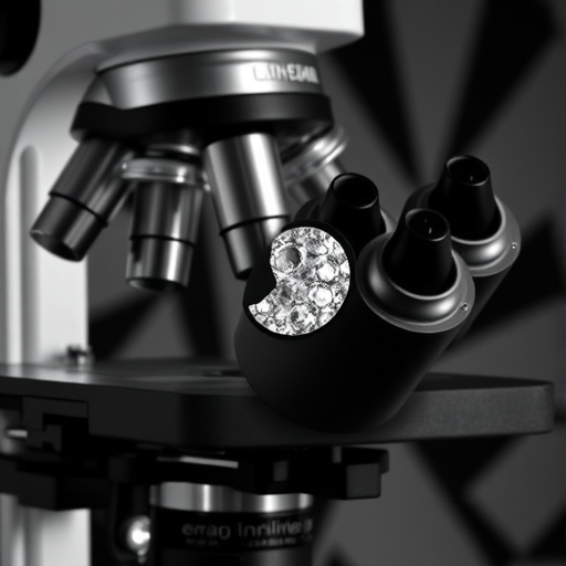
The innovative microscope, named PANORAMA, integrates a large telecentric photolithography lens, which was originally developed for high-precision chip manufacturing, combined with a substantial tube lens. This optical configuration projects the image onto an array of 48 small cameras arranged on a flat plane. Each individual camera captures a segment of the sample, and critically, each can be focused independently to accommodate variations in sample topography. This adaptive focusing ensures that even if the sample surface curves or tilts, every patch remains sharply in focus. By forgoing traditional scanning methods, PANORAMA achieves an imaging speed that was previously unattainable for such high-resolution large-area microscopy.
The technical mastery behind this system lies in its telecentric lens design. Unlike conventional lenses, telecentric lenses eliminate perspective distortion and maintain the same magnification regardless of object distance within a certain range. This characteristic enables the microscope to capture wide fields of view with minimal aberrations, an essential quality for stitching images from multiple cameras seamlessly. The lens system thus preserves the geometric fidelity of the sample across a substantial centimeter-scale imaging area while acquiring submicron resolution details, surpassing limitations inherent in single-sensor megapixel cameras.
By leveraging computational software, the images from all 48 camera modules are automatically stitched together to produce a continuous mosaic. This post-processing step, which takes roughly 5 to 10 minutes, assembles the segmented images into a seamless gigapixel-scale photograph characterized by astonishing detail. To contextualize the magnitude of this dataset, the resulting images contain 10 to 50 times more pixels than those taken by an average smartphone camera. This computational integration effectively flattens out the natural curvature of the sample surface, yielding a crisp focus throughout the entire field without mechanical stage movement.
Demonstrating the microscope’s capabilities, the team imaged a slide of rat brain tissue illuminated under brightfield conditions, capturing a 630-megapixel image in a single snapshot. Details as fine as 0.84 micrometers were resolvable, enabling visualization of neurons and dendritic structures across the sample. Such resolution corresponds to features approximately one-sixtieth to one-hundred-twentieth the diameter of a human hair. These findings underscore the microscope’s capacity for cellular-level imaging over expanses traditionally challenging for high-resolution systems.
The researchers further validated their setup by simultaneously acquiring brightfield and fluorescence images of onion skin positioned on a gently curved surface. Each camera module’s focus was individually adjusted to the local curvature, resulting in uniformly sharp images irrespective of the curvature. Brightfield images revealed distinct cell walls, while fluorescence imaging highlighted stained nuclei with high contrast. This dual-modality imaging exemplifies the microscope’s versatility for complex biological samples, potentially accelerating investigations in cytology and histology.
PANORAMA’s design eliminates the mechanical focusing and scanning that typically render gigapixel microscopy laborious and slow. Existing multi-camera microscopes often require stitching from multiple scans and manual refocusing steps, which can take up to an hour depending on sample size. In contrast, this single-shot methodology enables fast acquisition without compromising on image continuity or resolution. The absence of moving parts enhances robustness and reduces wear, making PANORAMA suitable for both research environments and industrial applications where throughput and reliability are critical.
Looking beyond current capabilities, ongoing development efforts are directed at scaling the field of view even further by increasing the number of cameras or employing larger sensors. Future iterations may capture entire petri dishes or sizable industrial surfaces with a comparable level of detail in a single shot. Additionally, automated focusing mechanisms are in progress to remove the need for manual adjustments, enhancing usability and efficiency. Advanced computational algorithms also hold promise for three-dimensional reconstructions and real-time imaging, potentially transforming the microscope into a dynamic tool for live cellular processes that evolve over time.
The researchers’ work represents a notable fusion of optical engineering and computational imaging strategies, setting a new paradigm in microscopy. Not only does it resolve classical trade-offs between field of view and resolution, but it also introduces an adaptive curvature compensation that was previously unattainable in gigapixel microscopy. The practicality and scalability of PANORAMA open new avenues across biomedical research, diagnostics, materials science, and manufacturing quality control, wherever detailed inspection of large or irregularly shaped specimens is required.
As this technology matures, it holds the potential to streamline workflows in pathology labs by enabling instant scanning of entire biopsy slides at cellular resolution, thus accelerating diagnosis and treatment decisions. Industrial inspectors might employ it to rapidly assess chip wafers or flexible materials with unprecedented granularity, preventing defects before they propagate. In research contexts, capturing expansive neural networks or plant tissues in their natural, non-flat state becomes vastly more achievable, empowering discoveries in biology and medicine.
This pioneering system exemplifies how marrying state-of-the-art optics with computational power can surmount fundamental limitations imposed by sample geometry and sensor size. By adapting the focus across individual camera modules in concert with a telecentric optical system, the microscope achieves uniform sharpness over curved surfaces without the need for mechanical intervention. Such innovations underline the transformative impact of interdisciplinary approaches in imaging science, driving advancements that ripple through science and technology sectors worldwide.
The publication detailing this breakthrough—“Curvature-adaptive gigapixel microscopy at submicron resolution and centimeter scale”—appeared in the journal Optics Letters on September 17, 2025. Given its potential, PANORAMA is expected to catalyze a wave of research and application, rendering previously arduous imaging tasks straightforward and rapid. As the technology evolves, enhancements in speed, automation, and multidimensional imaging are anticipated to solidify the system as a staple in advanced microscopy toolkits.
Subject of Research: Development of an adaptive multi-camera microscope for high-resolution gigapixel imaging of curved and large samples.
Article Title: Curvature-adaptive gigapixel microscopy at submicron resolution and centimeter scale
News Publication Date: 17-Sep-2025
Web References:
DOI link to article
Optica Publishing Group
Duke University
References:
X. Yang, H. Chen, L. Kreiss, C.B. Cook, G. Kuczewski, M. Harfouche, M.O. Bohlen, R. Horstmeyer, “Curvature-adaptive gigapixel microscopy at submicron resolution and centimeter scale,” Opt. Lett., 50, (2025). DOI: 10.1364/OL.572466
Image Credits: Xi Yang, Duke University
Keywords: Imaging, High resolution imaging, Medical imaging
Tags: biological tissue visualizationcurved sample imagingDuke University microscope innovationflexible materials imaginghigh-resolution microscopyimaging technology breakthroughsindustrial inspection microscopymedical diagnostics advancementsmulti-camera optical systemsnon-flat object imagingwide-angle imaging technology

