In a groundbreaking study published in the International Journal of Legal Medicine, researchers have unveiled new insights into sexual dimorphism within the human lumbar spine, using cutting-edge three-dimensional multi-slice computed tomography (MSCT) scanning techniques. This pioneering investigation explores the nuanced anatomical differences between male and female lumbar vertebrae, contributing significantly to forensic science, anthropological research, and clinical diagnostics. By employing advanced 3D imaging technologies, the team has offered a highly detailed morphometric analysis that promises to enhance identification protocols and foster deeper understanding of human skeletal variation.
Sexual dimorphism—the systematic difference in form between individuals of different sex in the same species—has long been a subject of intense scrutiny. However, traditional methods of assessing dimorphism have often relied on fragmented skeletal elements or two-dimensional imaging modalities, which lack the precision needed for comprehensive analysis. This new approach leverages multi-slice CT scans, which produce highly accurate and volumetric reconstructions of osseous structures, enabling an unprecedented level of detail when studying vertebral morphology.
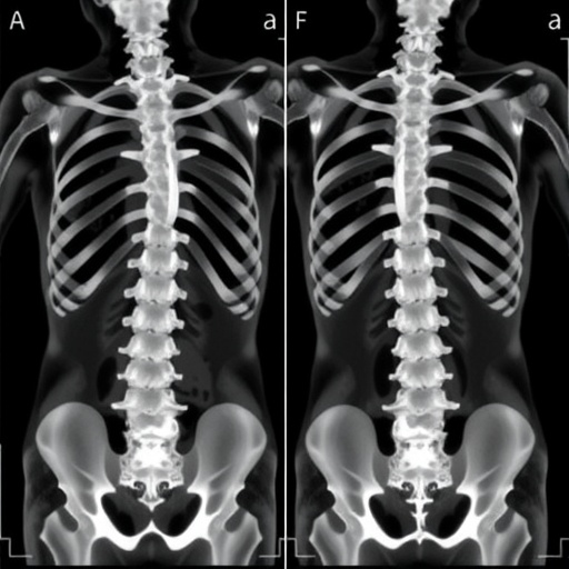
The lumbar vertebrae, comprising five large, robust bones situated in the lower back, play a crucial role in weight bearing and locomotion while offering protection to the spinal cord. Their structural characteristics often vary between males and females, but previous investigations rarely analyzed the lumbar column as a whole unit. This study stands out by meticulously evaluating every lumbar vertebra from L1 to L5, ensuring a full-spectrum comparative assessment that addresses individual vertebral variation and combined biomechanical function.
In this research, the scientists utilized MSCT scans sourced from a diverse cohort to generate three-dimensional models of lumbar vertebrae. These reconstructions allowed volumetric and morphometric measurements across multiple anatomical landmarks, including vertebral body height, width, depth, pedicle dimensions, and spinous process angles. The application of computational morphometrics further strengthened the statistical evaluation of sexual dimorphism by accommodating complex geometric variations rather than relying solely on linear measurements.
One of the most compelling outcomes is the identification of consistent, measurable differences across the entire lumbar spine that effectively distinguish males from females. For example, male lumbar vertebrae were found to be significantly larger in overall volume, exhibiting broader vertebral bodies and thicker pedicles. Conversely, female vertebrae showed comparatively smaller dimensions yet demonstrated less variation in certain morphological attributes, hinting at subtle biomechanical adaptations related to uniform load distribution.
These findings carry profound implications for forensic anthropology, where determining the sex of skeletal remains is often crucial but complicated by incomplete bones. Being able to analyze an entire lumbar series with high precision opens new avenues for identification, particularly in cases where other parts of the skeleton are unavailable or degraded. This holistic vertebral analysis could complement existing methods to increase accuracy in sex estimation, which underpins much of biological profiling in forensic contexts.
Beyond forensic applications, the study offers important clinical benefits, especially in the domain of spinal health and surgery. Understanding sex-specific lumbar vertebral anatomy is key to designing better implants, prosthetics, and surgical interventions tailored to anatomical variation. Moreover, this research may contribute to elucidating different susceptibilities to lumbar spine pathologies between males and females, such as disc degeneration, spondylolisthesis, or osteoporosis-related fractures.
The technological aspect of employing three-dimensional MSCT scanning cannot be overstated. Traditional radiographs provide limited views and suffer from superimpositions, while earlier CT technologies lacked the resolution to capture minute anatomical differences effectively. MSCT enables rapid, non-invasive capture of high-resolution volumetric datasets allowing multiplanar and 3D reconstructions indispensable for detailed morphometric studies. The computational power applied here exemplifies the synergy of medical imaging and data analytics in modern anatomical research.
Additionally, this study highlights the importance of standardizing measurement protocols for the vertebral column, as earlier studies often reported conflicting or incomparable results due to methodological variability. By utilizing objective landmarks in 3D space and leveraging advanced software tools, the researchers set a new benchmark for morbidity and biometric consistency in skeletal analyses, ensuring replicability and cross-study comparability.
With an increasing focus on personalized medicine and forensic precision, the investigation into sexual dimorphism via lumbar vertebrae represents a vital leap forward. Beyond sex estimation, the detailed morphometric datasets generated in this study could serve as reference points for population-specific anatomical variations, evolutionary biology research, and ergonomic design tailored to diverse human skeletal types.
Future research avenues stemming from this study might involve exploring sexual dimorphism across different age groups, ethnicities, and pathological conditions to understand how these variables impact lumbar vertebral morphology. Furthermore, integrating soft tissue imaging and biomechanical simulation could provide functional insights correlating anatomical dimorphism with movement patterns, injury mechanisms, and adaptive responses.
In terms of forensic applications, this methodology offers promising prospects when applied to fragmented or incomplete skeletal remains recovered in various investigative scenarios, including mass disasters, archaeological excavations, and criminal cases. The capacity to reconstruct and analyze vertebrae in three dimensions enhances the toolkit available to forensic anthropologists, potentially expediting identification processes with greater confidence.
Moreover, the research establishes a crucial link between state-of-the-art imaging technology and classical anatomical inquiry. It exemplifies how merging technological advancements with age-old investigative questions creates novel pathways for discovery and practical application, thereby transforming the landscape of medical and forensic sciences.
Overall, this comprehensive assessment underscores the value of the lumbar spine as a rich, albeit underutilized, source of sexual dimorphism data. By seamlessly integrating MSCT imaging, geometric morphometrics, and rigorous statistical analysis, the authors have forged an innovative approach that addresses long-standing challenges in anatomical science, setting the stage for enhanced forensic identification and clinical understanding.
As imaging techniques continue to evolve, the future of anatomical research lies in such integrative, multi-dimensional approaches. The insights garnered from this study not only provide immediate practical applications but also invite further interdisciplinary collaboration across fields such as radiology, forensic science, orthopedics, and bioengineering, igniting a new era of precision anatomy founded on technological innovation.
This seminal work, therefore, represents more than a mere morphological assessment—it serves as a testament to the transformative power of advanced imaging in unlocking the subtle, yet profound, biological differences that define the male and female human form.
Subject of Research: Assessment of sexual dimorphism in lumbar vertebrae using three-dimensional multi-slice computed tomography
Article Title: Assessment of sexual dimorphism in all lumbar vertebrae using three-dimensional multi-slice computed tomography scan
Article References:
Abd Elghany, S.A., Sharif, A.F., Mohammed Yehia, A.Y. et al. Assessment of sexual dimorphism in all lumbar vertebrae using three-dimensional multi-slice computed tomography scan. Int J Legal Med (2025). https://doi.org/10.1007/s00414-025-03594-z
Tags: 3D CT scan technologyadvanced imaging techniques in medicineanatomical differences in vertebraeclinical diagnostics in anthropologyforensic science applicationsimpact on identification protocolslumbar spine sexual dimorphismmale and female skeletal variationmorphometric analysis of lumbar vertebraemulti-slice computed tomography benefitssystematic difference in human anatomyvertebral morphology studies

