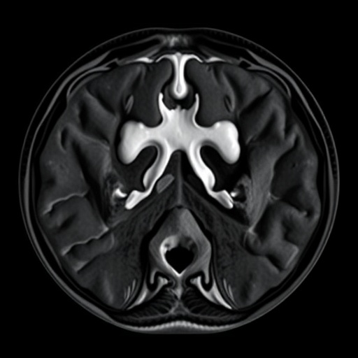
In a groundbreaking study poised to transform medical imaging, researchers have delved into the intricate relationship between deep learning technologies and the enhancement of synthetic high B-value diffusion-weighted imaging (DWI), specifically in the context of prostate lesion detection. This innovative research, conducted by Zhou, Zhang, Jiang, and colleagues, not only highlights advancements in imaging quality but also underscores the potential implications for patient diagnosis and treatment in the realm of urology.
The research tackles a pivotal concern in medical imaging: the quality of images obtained during MRI scans, which play a critical role in diagnosing various medical conditions. Prostate lesions, often challenging to detect and diagnose accurately, require a nuanced imaging approach to ensure that interpretations of these scans yield the necessary specificity and sensitivity. Traditional imaging techniques may fall short in providing the requisite clarity, thus propelling the need for innovative methods to enhance imaging results.
Deep learning, a subset of artificial intelligence, has emerged as a game-changer in various fields, including medical imaging. By utilizing sophisticated algorithms that mimic human cognitive processes, deep learning enables the analysis of vast amounts of data quickly, identifying patterns that may elude human observers. The potential for these technologies to improve image clarity and detail in medical images is an area of intense research and development.
In their recent publication, Zhou et al. present a novel approach that utilizes deep learning for the reconstruction of images from synthetic high B-value DWI. By employing advanced neural network architectures, the researchers have developed a method capable of generating high-quality magnetic resonance images that capture the subtle characteristics of prostate lesions more accurately than conventional methods. This improvement not only aids radiologists but also significantly impacts the clinical decision-making process.
The implications of enhanced imaging through deep learning are vast. A better understanding of the prostate’s internal landscape can lead to earlier detection of malignant lesions, potentially transforming patient outcomes. It is crucial for physicians to have access to the most accurate images possible, as these images serve as foundational pieces in developing effective treatment plans.
Another critical aspect of this research is the focus on synthetic high B-value DWI, which is a specific imaging technique that accentuates the contrast in tissue properties. This method is particularly beneficial for visualizing lesions that might be obscured at lower B-values. The challenge has been to ensure that the high B-value images do not introduce significant noise, which can compromise the clarity of the images and hinder accurate diagnosis.
Zhou and the team have tackled this issue head-on by integrating advanced deep learning techniques that refine the image reconstruction process, effectively reducing noise levels while enhancing image clarity. The result is a series of images that not only retain essential diagnostic features but also significantly improve overall interpretability for healthcare professionals.
As the healthcare sector increasingly leans towards technology-driven solutions, the potential for deep learning in imaging applications is becoming more evident. The ability to harness artificial intelligence could streamline workflows in radiology departments and allow healthcare providers to focus on higher-level patient care. The operational efficiencies gained from employing these advanced techniques may allow for quicker diagnoses, critical in the fast-paced environments of clinical medicine.
Further validating their findings, Zhou et al. also emphasize the importance of training these deep learning models with extensive datasets, which allows for the improvement of accuracy and reliability over time. By utilizing a robust dataset from diverse cohorts, the model can generalize its findings across different patient populations, ensuring that the advancements made in imaging quality are widely applicable and beneficial.
Despite the promising results, the researchers acknowledge that the integration of deep learning into clinical practices will require thorough validation and testing. As with any new technology, understanding the limitations and ensuring robust clinical trials will be imperative before widespread adoption can take place. Recognizing these hurdles is a necessary step towards creating a trustworthy and reliable medical imaging landscape.
The study published in the Journal of Medical Biology Engineering stands as a testament to the transformative potential of deep learning in the medical field. As similar advancements continue to emerge, the future of medical diagnostics holds significant promise, paving the way for more accurate, timely, and effective patient care across various specialties.
In summary, the fusion of deep learning and medical imaging signifies a critical intersection in healthcare innovation, offering pathways to improved diagnostic capabilities. The outcomes of Zhou et al.’s research illuminate the road ahead, providing hope that such advancements can become standardized in clinical settings, fulfilling the ongoing necessity to enhance detection and treatment methodologies in oncology.
The exploration of deep learning’s capabilities in improving image quality lays a vital cornerstone in the continuous evolution of prostate cancer diagnostics, making it an exciting field of study for researchers, clinicians, and patients alike. As we look toward the future, the integration of these advanced imaging techniques promises to elevate the standards of care and improve the accuracy with which we approach complex medical challenges.
Strongly supported by novel algorithms and deep learning frameworks, this research serves not only as an academic milestone but also as a beacon of innovation in health technology, setting the stage for future explorations into artificial intelligence’s role in medicine.
In closing, Zhou et al. have made a significant contribution that is poised to redefine prostatic lesion diagnostics in the near future, underscoring the essential relationship between technology and healthcare advancements. The incorporation of deep learning into image reconstruction processes represents not just an incremental improvement, but a paradigm shift in how we visualize and understand complex biological structures, thereby enhancing clinical outcomes and fostering a more effective approach to patient care.
Subject of Research: Prostate lesion detection using deep learning imaging techniques.
Article Title: Deep Learning Imaging-Based Reconstruction Improved the Image Quality of Synthetic High B-Value DWI for Prostate Lesion Detecting.
Article References:
Zhou, X., Zhang, H., Jiang, S. et al. Deep Learning Imaging-Based Reconstruction Improved the Image Quality of Synthetic High B-Value DWI for Prostate Lesion Detecting.
J. Med. Biol. Eng. 45, 157–165 (2025). https://doi.org/10.1007/s40846-025-00937-4
Image Credits: AI Generated
DOI: https://doi.org/10.1007/s40846-025-00937-4
Keywords: Deep learning, MRI, prostate lesions, medical imaging, artificial intelligence, image quality.
Tags: algorithmic analysis in imagingartificial intelligence in healthcaredeep learning in medical imagingenhanced imaging techniques for lesionsimaging specificity and sensitivitymedical imaging challengesMRI scan quality improvementpatient diagnosis and treatment implicationsprostate cancer diagnosis technologyprostate lesion detectionsynthetic high B-value diffusion-weighted imagingurology imaging advancements


