In a remarkable leap forward for regenerative medicine, researchers from the University of Minnesota Twin Cities have unveiled a pioneering technique that merges the cutting-edge technologies of 3D printing, stem cell biology, and engineered lab-grown tissues. This innovative approach holds the potential to revolutionize treatments for spinal cord injuries, addressing one of the most devastating medical challenges: the irreparable damage to nerve cells that leads to paralysis.
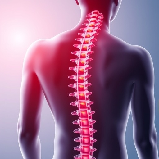
Spinal cord injuries, which affect over 300,000 individuals in the United States alone, have long posed an insurmountable barrier to complete recovery. The primary obstacle lies in the death of neurons at the injury site and the failure of severed nerve fibers to regenerate and reconnect. The new study, published in the prestigious journal Advanced Healthcare Materials, charts a novel path to overcoming these limitations through the use of 3D-printed organoid scaffolds that simulate the architecture of spinal tissue.
At the core of this breakthrough is a meticulously designed scaffold, produced through high-precision 3D printing. This framework incorporates microscopic channels structured to guide the growth and differentiation of spinal neural progenitor cells (sNPCs). These progenitor cells, which originate from human adult stem cells, possess the extraordinary ability to proliferate and mature into diverse types of neural cells necessary for reconstructing damaged spinal circuits.
.adsslot_Kh1noAkzE7{width:728px !important;height:90px !important;}
@media(max-width:1199px){ .adsslot_Kh1noAkzE7{width:468px !important;height:60px !important;}
}
@media(max-width:767px){ .adsslot_Kh1noAkzE7{width:320px !important;height:50px !important;}
}
ADVERTISEMENT
The scaffold’s microchannels serve not merely as a physical matrix but as directional conduits that instruct the regrowth of nerve fibers. By controlling the orientation and extension of new axons, the scaffold enforces a biologically relevant pattern of regeneration that aligns with the spinal cord’s natural connectivity. Guebum Han, the first author and a former postdoctoral researcher at the University of Minnesota, elaborates that this system acts akin to a neurobiological “relay,” effectively bypassing lesion sites to restore communication pathways.
The team evaluated the efficacy of their organoid scaffold by implanting it into a rat spinal cord model where the spinal cord was fully severed. The results were compelling: transplanted progenitor cells differentiated efficiently into neurons that extended axons bifurcating in both rostral and caudal directions. This bidirectional growth allowed the newly formed neural networks to establish functional synapses with the host’s existing spinal circuits, a critical step toward restoring motor and sensory function.
Crucially, longitudinal observations demonstrated that the new neural tissue integrated seamlessly over time with the host spinal cord, not eliciting significant immune rejection or scar tissue formation—common hurdles in neural regeneration therapies. The structural compatibility facilitated recovery of locomotor functions in treated rats, attesting to the therapeutic promise of this approach.
Ann Parr, a neurosurgery professor at the University of Minnesota and co-author of the study, highlights the transformational aspect of this research. She notes that regenerative medicine is entering an era where “mini spinal cords” grown ex vivo and fashioned into patient-specific implants could become feasible clinical interventions. The scaffold essentially resurrects the intrinsic regenerative capacity of the spinal cord by providing a conducive environment for cell growth and targeted axonal guidance.
Beyond the biological sophistication, this research introduces a scalable fabrication platform. The convergence of additive manufacturing with stem cell technology allows for customizable scaffolds that can be adapted to different injury geometries and patient-specific conditions. Such flexibility is vital for translating these findings from animal models to human clinical trials, where injury heterogeneity is substantial.
While still in early stages, the implications of these findings extend far beyond spinal cord injury treatment. The integration of 3D printed biomimetic scaffolds with progenitor cell populations may open future avenues in repairing other complex nervous system injuries and degenerative diseases. This convergence exemplifies the promise of interdisciplinary innovation at the nexus of engineering and biology.
The researchers plan to refine the scaffold’s design further, optimizing channel architecture and cell seeding protocols to enhance functional recovery. Parallel efforts will focus on ensuring long-term safety and efficacy, as well as developing good manufacturing practice (GMP)-compliant processes for clinical-grade scaffold production.
Funding from the National Institutes of Health, the State of Minnesota’s Spinal Cord Injury and Traumatic Brain Injury Research Grant Program, and the Spinal Cord Society has been instrumental in propelling this ambitious project. Collaborative expertise spanning mechanical engineering, neurosurgery, neuroscience, and physics has underpinned the robust translational strategy inherent in this work.
In summary, the University of Minnesota team’s advancement offers a beacon of hope for those affected by debilitating spinal cord injuries. By bridging bioengineering precision with stem cell biology, this research redefines regenerative paradigms and sets a new standard for restoring connectivity in damaged neural tissues. The full detailed findings and methodology are accessible through Advanced Healthcare Materials for the scientific community eager to build upon this foundation.
Subject of Research: Spinal cord injury recovery through 3D-printed organoid scaffolds incorporating spinal neural progenitor cells.
Article Title: 3D-Printed Scaffolds Promote Enhanced Spinal Organoid Formation for Use in Spinal Cord Injury
News Publication Date: 25 August 2025
Web References:
https://advanced.onlinelibrary.wiley.com/doi/10.1002/adhm.202404817
References:
The detailed study published in Advanced Healthcare Materials, DOI: 10.1002/adhm.202404817
Image Credits: McAlpine Research Group, University of Minnesota
Keywords: Spinal cord injuries, Organoids, Additive manufacturing, Stem cells, Tissue engineering
Tags: 3D-printed scaffolds for spinal cord injuriesadvanced healthcare materials researchcutting-edge medical technologiesengineered lab-grown tissuesneuron regeneration techniquesovercoming paralysis challengesregenerative medicine advancementsspinal cord injury treatment innovationsspinal neural progenitor cellsstem cell therapy for nerve repairtissue engineering for spinal recoveryUniversity of Minnesota spinal research

