
Reston, VA (August 22, 2025) — The field of nuclear medicine continually pushes the boundaries of diagnostic and therapeutic precision, and recent groundbreaking studies published ahead of print in The Journal of Nuclear Medicine (JNM) exemplify this innovative momentum. As the flagship publication of the Society of Nuclear Medicine and Molecular Imaging (SNMMI), these new research articles illuminate the evolving landscape of molecular imaging, precision diagnostics, and targeted radiotherapy, which together promise to revolutionize patient care through enhanced detection capabilities and novel treatment strategies.
One of the most promising advancements highlighted in the current issue is the pioneering work on positronium lifetime tomography. This cutting-edge imaging technique leverages the properties of positronium — a short-lived, hydrogen-like atom formed by an electron and its antiparticle, the positron — to delve deeper into tissue characterization than traditional imaging methods allow. By employing specialized scanners and radioactive tracers, scientists have succeeded in capturing minute variations in positronium lifetimes within living tissues. This allows for the construction of unprecedentedly detailed three-dimensional images with millimeter-level resolution, representing a significant leap toward sharper and more informative medical scans. The potential clinical impact is profound, especially in identifying subtle pathological changes that conventional modalities might overlook.
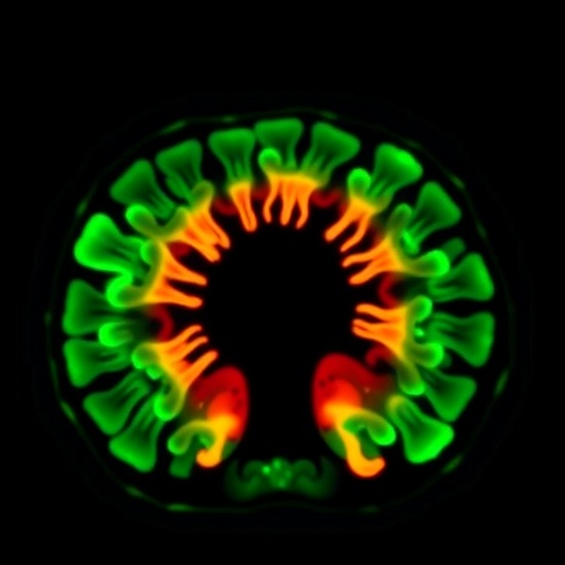
In parallel, researchers have made strides in oncology through the identification of new imaging biomarkers for endometrial cancer, a malignancy with rising incidence globally. Three biomarkers — HER2, MUC16, and CD24 — were investigated for their suitability as targets in positron emission tomography (PET) imaging. Using bespoke PET probes developed for these molecular targets, preclinical evaluation in cell cultures and murine models demonstrated strong tumor affinity for probes targeting HER2 and CD24. This heralds a new era in non-invasive tumor visualization that may improve early diagnosis, monitor therapeutic response, and ultimately personalize cancer treatment strategies. The incorporation of these novel biomarkers into routine clinical practice could significantly enhance the precision of endometrial cancer management.
.adsslot_XUaAHPWnEO{width:728px !important;height:90px !important;}
@media(max-width:1199px){ .adsslot_XUaAHPWnEO{width:468px !important;height:60px !important;}
}
@media(max-width:767px){ .adsslot_XUaAHPWnEO{width:320px !important;height:50px !important;}
}
ADVERTISEMENT
Another study addresses the formidable challenges in diagnosing Cushing disease, a hormonal disorder often caused by minuscule pituitary adenomas that evade detection with conventional magnetic resonance imaging (MRI). This multicenter cohort study evaluated the efficacy of combining MRI with carbon-11-labeled methionine PET (^11C-methionine PET/MRI) to improve tumor localization prior to surgical intervention. The dual-modality imaging demonstrated superior accuracy in identifying minute pituitary lesions, which is critical for surgical planning and optimizing patient outcomes. Such advancements underscore the power of hybrid imaging techniques in refining the diagnostic process for endocrine disorders traditionally considered elusive.
Beyond diagnostics, there are encouraging developments on the therapeutic front for sarcoma, a heterogeneous group of malignant tumors notorious for limited treatment options. Researchers explored the application of a novel targeted radiotherapy agent, ^177Lu-DOTAGA.Glu.(FAPi)2, designed to selectively bind to fibroblast activation protein (FAP) expressed in tumor stroma. In an initial clinical study involving ten patients who underwent multiple treatment cycles, the therapy was generally well tolerated, demonstrating manageable safety profiles. Imaging post-treatment revealed promising signs of tumor control in a subset of patients, suggesting that this radio-ligand therapy may evolve into a valuable option for managing difficult-to-treat sarcomas. This approach exemplifies the burgeoning field of theranostics — blending therapeutic and diagnostic capabilities within a single agent to optimize cancer care.
Taken together, these studies reflect a broader paradigm shift in nuclear medicine and molecular imaging toward highly individualized medicine, where diagnostic tools not only reveal disease presence but also provide intricate details tailored to individual pathology. The utilization of novel radiotracers, advanced detection technology, and hybrid imaging platforms facilitates this transformation, enabling clinicians to intervene earlier and with greater precision.
The advent of positronium lifetime tomography, for instance, may eventually allow clinicians to distinguish between benign and malignant lesions or detect microenvironmental changes associated with disease progression before structural anomalies manifest. Its nanosecond-scale temporal resolution, achieved through state-of-the-art detectors, opens unprecedented windows into physiological processes at the molecular level. Moreover, the scalability to whole-body imaging positions this technique as a versatile tool across multiple disease states.
Conversely, the incorporation of specific molecular biomarkers such as HER2 and CD24 into PET imaging algorithms represents a leap forward in tumor phenotyping. Their heterogeneous expression patterns and functional roles in cancer biology render them ideal candidates for targeted imaging probes, promising enhanced sensitivity and specificity. This improved molecular insight may pave the way for adaptive therapies, monitoring resistance mechanisms and guiding therapeutic switches in real-time.
In tackling pituitary adenomas responsible for Cushing disease, combining ^11C-methionine PET with high-resolution anatomical MRI epitomizes the integration of functional and structural data to resolve clinical dilemmas. Methionine, an essential amino acid, preferentially accumulates in active tumor tissue, augmenting contrast against surrounding normal structures. This synergy is crucial when dealing with lesions as small as a few millimeters, typically elusive to conventional imaging, thus refining surgical approaches to improve cure rates and reduce morbidity.
The exploration of ^177Lu-labeled FAP inhibitors for sarcoma therapy exemplifies the promise of targeted radionuclide therapy to selectively irradiate tumor microenvironments. FAP expression by cancer-associated fibroblasts is gaining recognition as a universal marker of tumor stroma, and targeting this niche could circumvent resistance mechanisms associated with tumor heterogeneity. Preliminary clinical data indicating safety and efficacy support further expansion of this therapeutic avenue.
Collectively, these innovative investigations illustrate the interdisciplinary collaboration across physics, chemistry, biology, and clinical medicine driving nuclear medicine into a new era. Enhanced by advances in radiochemistry, detector technology, and computational modeling, the field steadily approaches the ideal of highly sensitive, specific, and personalized diagnostic and therapeutic solutions.
The impressive range of current developments showcased in The Journal of Nuclear Medicine signals exciting times ahead for patients and healthcare providers alike. As these technologies mature and integrate into clinical practice, they hold the potential not only to transform diagnostic accuracy and therapeutic efficacy but also to fundamentally reshape clinical workflows and patient experiences in nuclear medicine.
For more in-depth information and access to the full research articles, visit The Journal of Nuclear Medicine online and follow their updates on social media platforms including Twitter, Facebook, and LinkedIn.
Subject of Research: Advances in molecular imaging, novel PET imaging biomarkers for endometrial cancer, positronium lifetime tomography, enhanced detection of Cushing disease, and targeted radiotherapy for sarcoma.
Article Title: Various—see individual study DOIs within The Journal of Nuclear Medicine.
News Publication Date: August 22, 2025
Web References:
Journal of Nuclear Medicine website: https://jnm.snmjournals.org/
Positronium lifetime tomography article: https://doi.org/10.2967/jnumed.125.270130
Endometrial cancer imaging article: https://doi.org/10.2967/jnumed.125.270318
Cushing disease imaging article: https://doi.org/10.2967/jnumed.124.269392
Sarcoma therapy article: https://doi.org/10.2967/jnumed.125.270186
Keywords: Molecular imaging, medical imaging, positron emission tomography, theranostics, precision medicine, targeted radiotherapy, endometrial cancer, Cushing disease, sarcoma, positronium lifetime tomography, PET/MRI, ^177Lu-DOTAGA.Glu.(FAPi)2
Tags: August 2025 journal highlightscutting-edge imaging techniquesdetailed tissue characterization methodsenhanced patient care strategiesmolecular imaging breakthroughsnuclear medicine advancementspositronium lifetime tomographyprecision diagnostics innovationsrevolutionary diagnostic toolsSociety of Nuclear Medicine and Molecular Imagingtargeted radiotherapy developmentsthree-dimensional medical imaging

