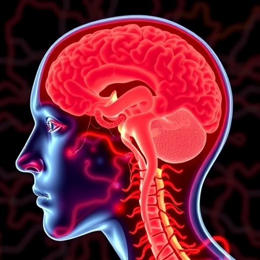
In a groundbreaking new study published in npj Parkinson’s Disease, researchers have shed light on the delicate interplay between cerebrovascular disease and cholinergic white matter pathways in the progression of Lewy body disorders. This investigation offers a pivotal advancement in understanding how vascular pathology affects neural networks integral to cognition and motor function in the Lewy body continuum, potentially opening new avenues for therapeutic intervention.
Lewy body diseases, encompassing Parkinson’s disease and dementia with Lewy bodies, are notoriously complex neurodegenerative disorders characterized by abnormal protein deposits called Lewy bodies. These pathologies disrupt neuronal communication, prominently within cholinergic circuits, which orchestrate attention, memory, and executive function. Prior research has hinted at vascular contributions to cognitive decline in these disorders, but a comprehensive, region-specific analysis connecting cerebrovascular pathology with white matter integrity remained elusive until now.
This study adopts an integrative neuroimaging approach to dissect the topographical relationships between cerebrovascular disease markers and cholinergic white matter pathways. By harnessing advanced diffusion tensor imaging alongside vascular burden assessments, the research team mapped how regional cerebrovascular insult correlates with microstructural degeneration in cholinergic tracts. Their findings underscore a non-uniform vulnerability of the cholinergic system to vascular compromise, intricately tied to Lewy body pathology stages.
.adsslot_AnkBVy1wqz{ width:728px !important; height:90px !important; }
@media (max-width:1199px) { .adsslot_AnkBVy1wqz{ width:468px !important; height:60px !important; } }
@media (max-width:767px) { .adsslot_AnkBVy1wqz{ width:320px !important; height:50px !important; } }
ADVERTISEMENT
What emerges from their data is a nuanced portrait of regional specificity. Certain cholinergic fiber bundles demonstrate heightened susceptibility to ischemic damage, which in turn exacerbates network disintegration. This selective pattern implies that vascular insults may accelerate or potentiate cholinergic failure in key cortical and subcortical circuits, thus enhancing symptom severity and clinical heterogeneity observed in patients with Lewy body disorders.
The implications of such regional associations are profound. By delineating which cholinergic pathways are most affected by cerebrovascular disease, clinicians and researchers can better predict the trajectory of cognitive decline and tailor interventions accordingly. For example, therapeutic strategies aimed at protecting or restoring blood flow in vulnerable white matter regions could mitigate cholinergic degradation and preserve neurological function.
Moreover, this work compels a reconsideration of the pathophysiological models underlying Lewy body diseases. Rather than viewing neurodegeneration in isolation, it emphasizes a pathological synergy where vascular and proteinopathies dynamically interact. Such an integrative perspective highlights the importance of addressing cerebrovascular health proactively in patients at risk of or diagnosed with Lewy body-related disorders.
The methodology employed in this study is particularly noteworthy. Leveraging diffusion MRI to assess white matter integrity allows a sensitive detection of microstructural changes not visible on conventional imaging. Combined with rigorous quantification of cerebrovascular burden, including white matter hyperintensities and lacunar infarcts, the study offers a robust framework for exploring neurovascular interactions in vivo.
In clinical terms, these findings could inform the development of novel diagnostic markers. Imaging signatures reflecting combined vascular and cholinergic pathway impairments might serve as early indicators of disease progression or treatment response. This is critical, given the pressing need for biomarkers in Lewy body disorders, which currently rely heavily on clinical observation and post-mortem confirmation.
Additionally, the regional vulnerability uncovered here aligns with known cognitive deficits in Lewy body disease. For instance, disruption of cholinergic pathways supplying the frontal cortex is strongly linked to executive dysfunction, a hallmark of dementia with Lewy bodies. Vascular damage in these areas may thus compound cognitive impairment, suggesting a mechanistic pathway for the clinical variability seen across patients.
Importantly, this study also challenges the previously held notion that cerebrovascular disease is merely a co-morbid condition in Lewy body disorders, instead positioning it as a crucial contributor to disease progression. This paradigm shift invites a broader clinical focus on vascular risk factors, such as hypertension and diabetes, as modifiable contributors to neurodegeneration in these populations.
The research team advocates for longitudinal studies to track the temporal interplay between vascular damage and cholinergic degeneration. Such investigations could illuminate causal relationships and potentially reveal windows for intervention before irreversible neuronal loss occurs. Understanding this chronology is vital for designing preventative strategies and improving patient outcomes.
From a therapeutic standpoint, the findings may rejuvenate interest in cholinesterase inhibitors and vascular protective agents. By integrating vascular risk management into standard care, alongside neuroprotective therapies, clinicians might slow cognitive decline more effectively. This dual-targeted approach could herald a new era in personalized medicine for Lewy body diseases.
Beyond clinical implications, this study adds a critical piece to the intricate puzzle of brain connectivity and resilience. The brain’s cholinergic networks are central to cognitive flexibility, and their vulnerability underscores the fragile balance between vascular supply and neural integrity. Recognizing how cerebrovascular disease disrupts this balance enriches our fundamental understanding of neurodegeneration.
In sum, the research by Rennie and colleagues crystallizes a complex, region-specific relationship between cerebrovascular pathology and cholinergic white matter damage within the Lewy body continuum. This finding compels the scientific and medical community to integrate vascular health more fully into the conceptual framework of Lewy body diseases, paving the way for innovative approaches to diagnosis, monitoring, and therapy.
This leap forward exemplifies how multidisciplinary neuroimaging and neuropathological investigation can uncover hidden dimensions of brain disease. As we delve deeper into the intertwined mechanisms of neurodegeneration and vascular dysfunction, the hope is to translate these insights rapidly into clinical tools that improve the quality of life for millions affected by these devastating disorders worldwide.
Subject of Research: Regional associations between cerebrovascular disease and cholinergic white matter pathways in the Lewy body continuum
Article Title: Regional associations between cerebrovascular disease and cholinergic white matter pathways in the Lewy body continuum
Article References:
Rennie, A., Nemy, M., Jerele, C. et al. Regional associations between cerebrovascular disease and cholinergic white matter pathways in the Lewy body continuum. npj Parkinsons Dis. 11, 250 (2025). https://doi.org/10.1038/s41531-025-01118-5
Image Credits: AI Generated
Tags: cerebrovascular disease and Lewy body disorderscholinergic circuits and executive functioncholinergic pathways in neurodegenerationcognitive decline in Lewy body diseasesdiffusion tensor imaging and cerebrovascular markersmicrostructural degeneration in cholinergic tractsneuroimaging in cholinergic researchParkinson’s disease and vascular healthregional cerebrovascular insult and neural networkstherapeutic interventions for Lewy body diseasesvascular contributions to cognitive deficitswhite matter integrity in neurodegeneration


