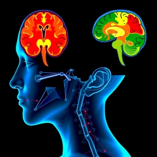
In a groundbreaking study published in Nature Neuroscience, researchers have unveiled compelling new insights into the remarkable stability of cortical body maps despite the traumatic event of arm amputation. This research challenges long-held assumptions about neural plasticity, showing that the brain’s primary body representation areas maintain their original functional organization even after the loss of a limb. These findings hold profound implications for our understanding of sensory processing, neurorehabilitation, and the persistent phantom sensations experienced by amputees.
For decades, neuroscientists have explored how the cortex adapts following injury, particularly in somatosensory regions responsible for representing the body’s surface. The somatosensory cortex, organized as a “body map,” allocates distinct spatial territories to different body parts. Classic theories posited that after an amputation, the cortical territory previously representing the lost limb would be rapidly taken over or “re-mapped” by adjacent body regions. Such plasticity was thought to underlie sensory compensation and changes in proprioception. However, this new work authored by Schone, Maimon-Mor, Kollamkulam, and colleagues presents strong evidence that these cortical representations remain remarkably stable pre- and post-amputation, challenging the notion of widespread cortical reorganization.
Using advanced functional magnetic resonance imaging (fMRI) techniques combined with sophisticated neural decoding algorithms, the authors meticulously mapped the detailed organization of somatosensory regions in human subjects both before and after arm amputation. Unlike prior studies that relied largely on cross-sectional data or animal models, this longitudinal design allowed for direct comparisons over time within the same individuals. Such precision afforded the team the ability to measure subtle functional changes with unprecedented fidelity.
.adsslot_1lMQHfX6z8{ width:728px !important; height:90px !important; }
@media (max-width:1199px) { .adsslot_1lMQHfX6z8{ width:468px !important; height:60px !important; } }
@media (max-width:767px) { .adsslot_1lMQHfX6z8{ width:320px !important; height:50px !important; } }
ADVERTISEMENT
The results revealed that, even years after amputation, the somatosensory cortex maintained a persistent topographic representation of the missing limb. Notably, the spatial patterning of neural activity evoked by imagined movements of the absent hand closely mirrored that of the intact limb or pre-amputation state. Such preservation suggests that the brain’s “body map” operates as a deeply entrenched neural scaffold resistant to structural changes triggered by sensory loss. This contradicts previous assumptions that neural circuits reorganize dramatically within the primary somatosensory cortex.
The study further delved into the mechanisms underpinning this stability by examining the connectivity profiles between somatosensory and motor cortical areas. Their findings suggest that the preserved cortical maps are functionally interconnected with effector regions controlling the residual limb and the contralateral intact body parts, forming a resilient network. This could explain why the cortical territory is not easily overwritten by adjacent body parts, contradicting prevailing notions of competitive neural territory plasticity.
Moreover, the authors correlated these neural findings with behavioral assessments, including tactile perception and phantom limb movement control. They discovered that individuals with stronger preserved cortical maps demonstrated enhanced ability to voluntarily modulate phantom limb movements, indicating that these stable representations maintain functional relevance beyond mere anatomical retention.
The implications of this work extend far beyond theoretical neuroscience. Understanding the immutable nature of cortical body maps opens new avenues for prosthetic limb control technologies that harness these pre-existing neural circuits. Brain-machine interfaces designed to decode these preserved neural signals could vastly improve the intuitiveness and precision of advanced prostheses, restoring meaningful motor and sensory experiences to amputees.
In addition, the revelation that somatosensory cortical maps remain unaltered despite limb loss prompts a reevaluation of neurorehabilitation strategies, many of which rely on inducing cortical plasticity to retrain the brain. Therapies might instead focus on leveraging stable cortical networks, substituting damaged sensory pathways with artificial feedback to optimize recovery and adaptation.
This research also raises fascinating questions about the developmental origins and robustness of cortical maps. The authors hypothesize that once established, these maps are deeply encoded within both cortical microcircuits and broader neural architecture, potentially informed by genetic and embryonic patterning cues. The degree of post-injury plasticity, therefore, may be far more limited than previously believed, at least in primary somatosensory areas.
Further studies will be necessary to explore whether similar stability exists in other sensory or motor cortical regions, such as those representing the lower limbs or facial features. Additionally, exploring the temporal dynamics of these representations immediately after injury versus years later could elucidate critical windows of plasticity or consolidation.
Intriguingly, these findings may also inform treatments of neurological conditions beyond amputation. For example, stable cortical maps could underlie the persistence of maladaptive sensory experiences in conditions like complex regional pain syndrome or stroke, pointing towards novel targets for intervention.
In summary, the meticulous work by Schone et al. presents a paradigm-shifting view that cortical body maps are remarkably stable neural substrates, resilient to even severe sensory deprivation caused by limb loss. This challenges entrenched beliefs about brain plasticity and opens exciting new horizons for neuroscience, neuroprosthetics, and clinical rehabilitation. The enduring cortical representation of the missing limb offers a window into the brain’s enigmatic capacity for stable self-representation amidst profound bodily change.
As the field advances, integrating these insights with cutting-edge neurotechnology could revolutionize the quality of life for millions of amputees worldwide. Harnessing the power of persistent cortical body maps may ultimately enable seamless mind-controlled prosthetics and novel therapies for phantom limb pain, transforming trauma into tangible neural opportunity. This research not only deepens fundamental understanding but brings hope for future innovations grounded in the brain’s extraordinary stability.
Subject of Research: Cortical body map stability and neural representation after arm amputation
Article Title: Stable cortical body maps before and after arm amputation
Article References:
Schone, H.R., Maimon-Mor, R.O., Kollamkulam, M. et al. Stable cortical body maps before and after arm amputation. Nat Neurosci (2025). https://doi.org/10.1038/s41593-025-02037-7
Image Credits: AI Generated
Tags: advanced fMRI techniques in neuroscience researchcortical body maps after arm amputationimplications for neurorehabilitation strategiesimplications of arm amputation on brain organizationinsights into brain injury and recovery processesneural plasticity and cortical stabilityphantom limb sensations in amputeesresearch on limb loss and brain adaptationsensory processing in the somatosensory cortexstability of body representation in the brainunderstanding cortical territory allocationunderstanding sensory compensation mechanisms


