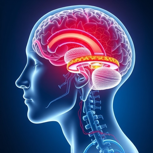
In a groundbreaking new study published in npj Parkinson’s Disease, researchers have unveiled the intricate mechanisms by which iron mishandling occurs both in the brain and peripheral systems of individuals afflicted with Parkinson’s disease. This exploration sheds new light on the complex biochemistry underlying the disease, offering promising avenues for future therapeutic interventions that could significantly alter the course of this debilitating neurodegenerative disorder. By dissecting the pathways of iron metabolism and its dysregulation, the study provides compelling evidence that iron imbalance is not merely a bystander but a central player in Parkinson’s pathology.
Parkinson’s disease, traditionally characterized by its hallmark motor symptoms such as tremors, rigidity, and bradykinesia, has long been linked to the degeneration of dopaminergic neurons in the substantia nigra of the brain. However, the precise molecular drivers of neuronal death have remained elusive. This research highlights that abnormal iron accumulation within specific brain regions exacerbates oxidative stress and fosters neuronal vulnerability. These findings dovetail with the growing consensus that metal ion homeostasis plays a pivotal role in neurodegeneration, intersecting with protein aggregation, mitochondrial dysfunction, and inflammatory processes.
The study methodically demonstrates that the mishandling of iron is not confined to the central nervous system but extends systemically, implicating peripheral tissues in the pathogenic cascade. Through meticulous biochemical assays and advanced imaging, the researchers quantified iron levels across multiple organ systems in Parkinson’s patients and matched controls. Their data reveal a distinctive pattern of iron dysregulation that mirrors the neuropathological features observed in the brain, underscoring the systemic nature of this metal’s toxic potential. This systemic disturbance challenges the existing focus solely on cerebral changes and prompts a reconsideration of Parkinson’s as a multi-organ disorder.
.adsslot_V4gNC3Qzda{ width:728px !important; height:90px !important; }
@media (max-width:1199px) { .adsslot_V4gNC3Qzda{ width:468px !important; height:60px !important; } }
@media (max-width:767px) { .adsslot_V4gNC3Qzda{ width:320px !important; height:50px !important; } }
ADVERTISEMENT
Iron homeostasis is maintained through a delicate equilibrium involving iron transport, storage, and regulatory proteins. In healthy states, ferritin sequesters excess iron safely, while transferrin mediates its circulation. The research elucidates profound disruptions in these systems among Parkinson’s patients. Elevated levels of free, reactive iron were detected in the substantia nigra and peripheral blood, indicative of impaired sequestration. Furthermore, proteins such as ferroportin and hepcidin, which govern iron export and absorption, displayed aberrant expression patterns. This dysregulated iron trafficking fosters an environment ripe for Fenton chemistry reactions, generating excessive reactive oxygen species that contribute to neuronal demise.
One of the most significant insights from this research is the demonstration of a feedback loop wherein oxidative stress intensifies iron mishandling, which in turn escalates oxidative damage. Neurons in Parkinson’s disease are especially susceptible to oxidative insults due to their high metabolic rate and relatively limited antioxidant defenses. The interplay between iron overload and oxidative stress creates a vicious cycle, accelerating neurodegeneration. These insights explain why certain neurons, particularly in the substantia nigra pars compacta, are preferentially vulnerable, correlating with the clinical manifestations of the disease.
Beyond iron’s direct involvement, the study ventures into the realm of peripheral immune responses and their connection with iron metabolism. Dysregulated iron availability influences immune cell function, notably in microglia and macrophages, shaping inflammatory responses that exacerbate neuronal injury. Peripheral immune cells bearing iron overload may contribute to systemic inflammation, thus amplifying neuroinflammation through the blood-brain barrier. These findings bridge the gap between central nervous system pathology and peripheral immune activation, which has been observed but poorly understood in Parkinson’s disease.
Moreover, the researchers employed cutting-edge imaging techniques to visualize iron deposits in vivo, advancing diagnostic capabilities. Magnetic resonance imaging (MRI) with iron-sensitive sequences revealed distinct iron accumulation patterns in Parkinson’s patients compared to controls. This non-invasive biomarker could pave the way for earlier diagnosis and monitoring disease progression, providing a vital tool for clinicians and researchers alike. Combined with biochemical markers of iron metabolism in peripheral blood, these imaging advances present a dual modality approach to track disease dynamics.
Therapeutically, the research sheds light on potential strategies targeting iron homeostasis to halt or slow Parkinson’s progression. Chelation therapies that bind free iron and reduce its availability have been studied but remain underutilized due to concerns about systemic iron depletion and side effects. This study suggests that more nuanced approaches aimed at restoring regulatory protein function or preventing iron-induced oxidative damage may be more beneficial. For instance, modulating hepcidin or ferroportin expression could recalibrate iron handling without inducing iron deficiency, offering a refined therapeutic window.
Another promising avenue involves enhancing the antioxidative defenses of neurons. By mitigating the oxidative stress resulting from iron mishandling, it may be possible to preserve neuronal integrity. Compounds that upregulate endogenous antioxidants or mimic their activity are being investigated in preclinical models. The concurrent targeting of iron dysregulation and oxidative stress represents a dual-pronged approach that could revolutionize Parkinson’s therapeutics, moving beyond symptomatic treatment toward disease modification.
Interestingly, this study also alludes to the genetic underpinnings influencing iron metabolism in Parkinson’s disease. Variants in genes encoding proteins responsible for iron handling may predispose individuals to iron accumulation and neurodegeneration. Genome-wide association studies have identified several such candidates, but functional validation remains scarce. This research highlights the importance of integrating genetic data with biochemical and imaging findings to unravel the heterogeneity of Parkinson’s disease and tailor personalized treatment strategies.
Environmental factors that alter systemic iron levels or promote iron accumulation in the brain are also considered. Exposure to iron-rich environments or dietary iron overload could exacerbate Parkinson’s pathology, although causality remains to be established. The interplay of genetics, environmental triggers, and systemic iron handling constructs a multifactorial framework for disease development, suggesting that interventions might need to address several axes simultaneously.
The implications of this research extend to other neurodegenerative diseases marked by iron accumulation, such as Alzheimer’s disease and multiple sclerosis. Cross-disease comparisons of iron mishandling mechanisms could uncover shared pathological pathways, facilitating broad-spectrum therapeutics. The fundamental insight that iron’s redox activity, while essential for cellular metabolism, becomes deleterious when misregulated resonates across the neurodegeneration field and reinforces the urgency of understanding metal biology.
In summary, this comprehensive investigation into iron’s dual pathology within the brain and peripheral tissues enriches our understanding of Parkinson’s disease and heralds a paradigm shift. By reframing the disease as a systemic disorder influenced by iron metabolism, it opens fertile ground for innovative diagnostics and therapeutics. The journey from molecular insight to clinical application, though demanding, promises hope for millions of Parkinson’s patients worldwide.
As we await further clinical trials that test iron modulation in Parkinson’s disease, this study serves as a clarion call to the scientific community to continue unraveling the complexities of metal homeostasis in neurodegeneration. The convergence of molecular biology, imaging technology, genetics, and pharmacology exemplifies the interdisciplinary approach needed to tackle the formidable challenge posed by Parkinson’s disease. Ultimately, the elucidation of iron mishandling propels us closer to deciphering the enigma of neurodegeneration.
Subject of Research: Iron metabolism dysregulation in Parkinson’s disease and its systemic effects
Article Title: Iron mishandling in the brain and periphery in Parkinson’s disease
Article References:
Bolen, M.L., Menees, K.B., Dupreez, A.C. et al. Iron mishandling in the brain and periphery in Parkinson’s disease.
npj Parkinsons Dis. 11, 246 (2025). https://doi.org/10.1038/s41531-025-01089-7
Image Credits: AI Generated
Tags: biochemistry of Parkinson’s diseasedopaminergic neuron degeneration mechanismsinflammatory processes in Parkinson’siron accumulation in brain regionsiron metabolism in Parkinson’s diseasemetal ion homeostasis in neurodegenerationmolecular drivers of neuronal deathneurodegenerative disorders and iron imbalanceoxidative stress and neuronal vulnerabilitypathways of iron dysregulationsystemic implications of iron mishandlingtherapeutic interventions for Parkinson’s


