In the relentless quest to visualize the intricate machinery of life at the molecular scale, microscopy techniques have continuously evolved to break through the barriers of optical resolution. Among the forefront technologies, MINFLUX microscopy has emerged as a revolutionary method, pushing the boundaries of fluorescence imaging into the realm of nanometer precision. By ingeniously combining patterned excitation with point-detection schemes, MINFLUX achieves molecular localization with exceptional spatial accuracy. However, its widespread adoption has been hampered by inherent technical challenges, notably the complexity involved in the iterative scanning process that it demands.
MINFLUX operates by focusing a structured laser excitation pattern onto fluorescent molecules and analyzing their emitted photons to pinpoint their positions. The method hinges upon sequentially scanning smaller and smaller areas, refining localization estimates progressively to achieve nanometric precision. This meticulous scanning regime, while effective, introduces a trade-off between acquisition speed and dimensional range, besides raising the bar for experimental setup complexity and ease of use. Essentially, although MINFLUX sets a new gold-standard for spatial resolution, it often limits throughput and accessibility for broader biological applications.
A recent breakthrough from the team led by Dr. Giuseppe Vicidomini at the Istituto Italiano di Tecnologia has now addressed these limitations through the development of ISM-FLUX—a robust, streamlined reimagining of the conventional MINFLUX paradigm. Published in Light: Science & Applications, this innovation leverages advances in detector technology to fundamentally simplify the localization process while retaining, and in some aspects enhancing, the exquisite nanoscale spatial resolution that defined the original method.
.adsslot_nModewtZrL{ width:728px !important; height:90px !important; }
@media (max-width:1199px) { .adsslot_nModewtZrL{ width:468px !important; height:60px !important; } }
@media (max-width:767px) { .adsslot_nModewtZrL{ width:320px !important; height:50px !important; } }
ADVERTISEMENT
At the heart of ISM-FLUX lies an asynchronous readout single-photon avalanche diode (SPAD) array detector. Unlike traditional MINFLUX systems, which rely on a single-element detector collecting integrated photon counts sequentially during laser scans, ISM-FLUX employs a 5×5 SPAD array. This detector array provides a rich spatiotemporal dataset by capturing the precise arrival time and position of individual fluorescence photons across the array, thereby encoding invaluable localization information that single-pixel detectors inherently discard.
This multidimensional data acquisition transforms the photonic detection landscape. By correlating the spatial coordinates of excitation beams with the spatially resolved photon detection events, ISM-FLUX realizes a dramatically expanded localization range. Instead of being confined to iterative scanning of progressively smaller regions, ISM-FLUX can localize molecules accurately over considerably larger fields without compromising the localization precision that defines MINFLUX, effectively blending the high resolution of the original technique with the expansive field advantages reminiscent of image scanning microscopy.
The implications for biological imaging are profound. Validation experiments utilizing fluorescent molecules and DNA origami nanorulers with fluorophores spaced a mere 6 nanometers apart demonstrated ISM-FLUX’s capability to resolve structures at a scale beyond conventional diffraction limits. This capability suggests that researchers can now observe and quantify molecular assemblies and interactions with unprecedented minuteness and clarity, in a fraction of the time previously required.
Notably, ISM-FLUX’s simplified methodology does more than enhance throughput; it marks a significant stride in democratizing advanced fluorescence nanoscopy. By removing the necessity for complex, multistep scanning and adopting readily integrable SPAD array detectors, the technology lowers both technical hurdles and operational demands. As Dr. Vicidomini’s team envisions, future implementations of this method could encourage universal integration of SPAD arrays across diverse laser-scanning microscopy platforms, expanding their molecular imaging capacity.
The principle behind the technology stems from innovating beyond photon counting to photon mapping, capturing not just the quantity but also the positional context of each emitted photon in real time. The 5×5 SPAD array detector operates with ultra-high timing resolution and negligible dark counts, attributes which are vital in detecting single-photon events with extreme sensitivity. This level of detection sensitivity married with spatial awareness transforms raw photon data into a multidimensional matrix ripe for computational reconstruction, paving the way for localization algorithms to exploit richer datasets.
Moreover, by forgoing the iterative miniaturization of scanned regions, ISM-FLUX sidesteps cumulative photobleaching effects and reduces phototoxicity risks on biological specimens—challenges that have historically constrained live-cell imaging in super-resolution studies. The ability to maintain high localization precision across relatively larger scanning fields implies less laser exposure per molecular target and faster acquisition of statistically meaningful images.
The transition from single-element detectors to multi-pixel, asynchronous photon detection arrays represents a broader paradigm shift in fluorescence microscopy, reflecting the intersection of photonics engineering, computational optics, and molecular biology. ISM-FLUX epitomizes this confluence, aligning detector innovation with algorithmic sophistication to yield a tool that promises to unlock new vistas in observing molecular dynamics within living cells with nanoscale resolution.
In summary, ISM-FLUX heralds an evolution in MINFLUX microscopy by combining spatially resolved single-photon detection with efficient localization algorithms, overcoming longstanding challenges of complexity and limited fields of view. This breakthrough not only expedites the path toward widespread adoption of molecular-scale imaging but also enriches the data quality and biological relevance of fluorescence microscopy. As the technology matures, its potential to illuminate molecular mechanisms underlying health and disease could catalyze transformative advancements in cell biology, neurobiology, and beyond.
The work epitomizes how leveraging state-of-the-art detector arrays can refine and simplify intricate optical techniques without sacrificing performance, offering a blueprint for future innovations in nanoscale optical imaging. It underlines the promising horizon where precision, speed, and simplicity coexist, empowering a wider community of researchers to explore the molecular complexity of life in real time.
Subject of Research:
Advanced fluorescence microscopy technique development for molecular-scale localization.
Article Title:
Array Detection Enables Large Localisation Range for Simple and Robust MINFLUX
Web References:
10.1038/s41377-025-01883-1
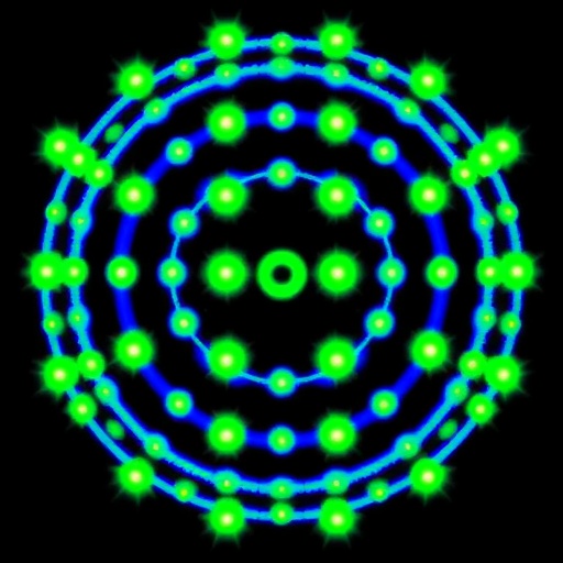
Image Credits:
Eli Slenders et al.
Keywords
MINFLUX, ISM-FLUX, Single-Photon Avalanche Diode (SPAD) Array, Fluorescence Microscopy, Super-Resolution Imaging, Nanometer Precision, Molecular Localization, DNA Nanostructures, Photon Detection, Laser Scanning Microscopy, Molecular Imaging Technologies, Optical Nanoscopy
Tags: acquisition speed versus dimensional rangebiological applications of MINFLUXbreakthroughs in microscopy technologyenhancing imaging accessibility and throughputexperimental setup complexity in microscopyfluorescence imaging advancementsiterative scanning process challengesMINFLUX microscopymolecular scale imaging techniquesnanometer precision localizationphotonic emission analysisstructured laser excitation patterns

