
In recent years, the scientific community has increasingly turned its attention to understanding the subtle yet profound impacts of preterm birth on brain development and cognition. A groundbreaking study published in Pediatric Research (2025) sheds new light on how children born moderate to late preterm (MLP) differ in their neurodevelopmental trajectories compared to their full-term peers. By employing state-of-the-art neuroimaging techniques, this research pushes forward the frontiers of knowledge regarding the lasting cognitive implications of MLP birth during late childhood.
The study zeroes in on the window of late childhood, a critical period characterized by intensive brain maturation and cognitive skill refinement. The authors, led by Acosta-Rodriguez et al., employed advanced magnetic resonance imaging (MRI) protocols to analyze structural and functional brain markers associated with cognition. Their interest was specifically targeted toward children born between 32 and 36 weeks of gestation, a group historically understudied relative to extremely preterm infants. This nuanced focus allows researchers to decipher the more subtle neurodevelopmental alterations that might not be evident in infancy but emerge with increasing cognitive demands.
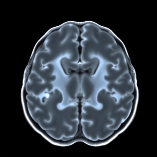
One of the most striking aspects of the study is its comprehensive approach combining cognitive assessments with high-resolution neuroimaging. The researchers utilized diffusion tensor imaging (DTI), resting-state functional MRI (rs-fMRI), and volumetric analyses to chart alterations in white matter integrity, connectivity patterns, and regional brain volumes. These multi-modal imaging strategies enable a holistic interpretation of the brain’s architecture and function, critical to understanding how premature birth influences neurological substrates that underpin learning and memory.
.adsslot_4K1vuSsane{width:728px !important;height:90px !important;}
@media(max-width:1199px){ .adsslot_4K1vuSsane{width:468px !important;height:60px !important;}
}
@media(max-width:767px){ .adsslot_4K1vuSsane{width:320px !important;height:50px !important;}
}
ADVERTISEMENT
The results elucidate a pattern of altered neurodevelopment in MLP children that, while less severe than in extremely preterm infants, nonetheless reveals significant disparities compared to full-term controls. Notably, the integrity of specific white matter tracts, such as the arcuate fasciculus and the superior longitudinal fasciculus—key pathways supporting language and executive function—showed reduced microstructural organization correlating with diminished performance in standardized cognitive tests. This finding highlights the importance of white matter maturation in the context of prematurity-related cognitive challenges.
Moreover, the study highlights intriguing variations in functional connectivity within networks implicated in attention and cognitive control. Resting-state fMRI revealed that MLP children exhibited less synchronized activity between prefrontal and parietal cortices, suggesting a potential neural basis for commonly reported attentional difficulties and executive dysfunction in this population. These disrupted network dynamics may reflect subtle delays in the maturation of neuronal circuits crucial for higher-order cognitive processes.
Simultaneously, volumetric analyses uncovered region-specific reductions in gray matter volume, particularly within the prefrontal cortex and hippocampus—structures integrally involved in working memory, decision-making, and learning. These morphological differences provide a tangible neural correlate for observed neuropsychological deficits, underscoring how even modest degrees of prematurity can affect cerebral growth trajectories.
A crucial component of this investigation is its emphasis on late childhood as a critical evaluation period. Previous research often concentrated on infancy or early childhood, potentially overlooking complications that consolidate or emerge later as cognitive demands increase. By focusing on a later developmental stage, this study reveals that neurodevelopmental vulnerabilities associated with MLP birth are not transient but persist, potentially influencing academic achievement and psychosocial adaptation during pivotal stages of child development.
The findings bear significant implications for medical and educational interventions. Early identification of neurodevelopmental divergence via neuroimaging could facilitate targeted support that caters to the specific cognitive domains affected in MLP children. This precision medicine approach might enable clinicians and educators to tailor therapies and learning environments that mitigate risks of long-term difficulties, thereby enhancing life trajectories.
Importantly, the study also recognizes the heterogeneity within the MLP group, underscoring that prematurity is not a monolithic risk factor. Variability in perinatal health, socioeconomic status, and postnatal experiences contribute to divergent neurocognitive outcomes. Future research may thus benefit from integrating genetic and environmental data to comprehensively map factors that modulate vulnerability and resilience following moderate to late preterm birth.
Methodologically, the research exemplifies the growing power of neuroimaging biomarkers as objective, quantifiable indices of brain health and cognition in pediatric populations. The fusion of structural, functional, and microstructural metrics offers a powerful framework for charting developmental trajectories and identifying atypical patterns that may predict neurodevelopmental disorders or learning disabilities well before clinical symptoms become evident.
The study’s revelations also open doors for longitudinal research trajectories. By tracking this cohort into adolescence and adulthood, scientists can map the evolution of brain-cognition relationships and determine critical periods where interventions may be most efficacious. Such longitudinal insights will be instrumental in unraveling the plasticity and recovery potential of the developing brain after early-life insult such as prematurity.
Beyond clinical and educational spheres, these neuroimaging findings contribute to fundamental neuroscience by illuminating how gestational age modulates the maturation of brain networks underpinning cognition. The observed alterations in white matter connectivity and cortical volumes elucidate biological pathways through which preterm birth disrupts normative brain maturation, advancing our grasp of neurodevelopmental processes at a systems level.
Intriguingly, this line of research prompts further investigation into potential neurobiological mechanisms driving these alterations, including disruptions in oligodendrocyte development, synaptic pruning, and neuroinflammation associated with perinatal stressors. Understanding these intracellular and molecular underpinnings is critical for devising therapeutic strategies that could promote optimal neural repair or plasticity during sensitive developmental windows.
Furthermore, the study invites a revisitation of how clinical definitions of prematurity incorporate neurodevelopmental risk. While extremely preterm birth is widely recognized for its adverse neurocognitive sequelae, moderate to late preterm birth accounts for a far larger portion of preterm deliveries and thus impacts public health on a broader scale. This research underscores the need for heightened surveillance and support for this broader cohort.
In the current era of precision pediatrics, such comprehensive neuroimaging work represents a paradigm shift from purely behavioral assessments toward personalized brain-based evaluations. These methodologies could ultimately refine diagnostic criteria, predict individual prognosis, and tailor interventions that holistically consider the neural bases of cognitive strengths and vulnerabilities.
As the scientific world digests these findings, the hope is that we move beyond viewing MLP birth as a benign clinical category. Instead, this research urges recognition of its latent but tangible effects on neurodevelopment, encouraging health providers, educators, and families to adopt a proactive stance in optimizing developmental outcomes for these children.
The study by Acosta-Rodriguez and colleagues, therefore, marks a pivotal contribution to contemporary pediatric neuroscience, leveraging cutting-edge neuroimaging to illuminate how relatively common early-life experiences shape the architecture and function of the growing brain. It signals a call to action: to recognize, understand, and address the subtle cognitive footprints left by moderate to late prematurity in pursuit of healthier futures.
Subject of Research: Neurodevelopmental associations and cognitive outcomes in children born moderate to late preterm (MLP) during late childhood.
Article Title: Neuroimaging markers of cognition in late childhood associated with moderate to late preterm birth.
Article References:
Acosta-Rodriguez, H., Bobba, P., Zeevi, T. et al. Neuroimaging markers of cognition in late childhood associated with moderate to late preterm birth. Pediatr Res (2025). https://doi.org/10.1038/s41390-025-04286-5
Image Credits: AI Generated
DOI: https://doi.org/10.1038/s41390-025-04286-5
Tags: advanced MRI protocols in pediatric studiesbrain development in preterm infantscognitive skills refinement in childhoodcognitive traits in moderate pretermsdiffusion tensor imaging in cognitive assessmentsimpact of preterm birth on cognitionlate childhood neurodevelopmentmoderate to late preterm birth outcomesneurodevelopmental trajectories of preterm childrenneuroimaging techniques in cognitive researchpediatric neurodevelopmental research findingsstructural and functional brain markers


