A groundbreaking advancement in the realm of artificial intelligence (AI) is set to revolutionize the medical imaging landscape. Researchers at the University of California San Diego have developed a new AI tool that significantly simplifies and reduces the cost associated with training medical imaging software. This innovation is especially beneficial when the number of available patient scans is limited, addressing a persistent challenge in the healthcare field.
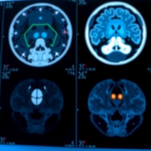
Medical image segmentation, the core focus of this breakthrough, involves labeling each pixel in an image according to its characteristic—distinguishing between cancerous tissue and healthy tissue, for instance. Currently, this meticulous task is predominantly performed by expert radiologists or trained specialists, as deep learning techniques have shown potential to assist in automating this process. However, these methods traditionally depend heavily on access to vast datasets comprising pixel-by-pixel annotated images.
The necessity for extensive annotated datasets poses a significant hurdle for the implementation of deep learning techniques in medical contexts. Li Zhang, a Ph.D. student within the Department of Electrical and Computer Engineering at UC San Diego, explains that compiling such datasets can be a labor-intensive endeavor. This process demands considerable time, expertise, and financial resources, often resulting in a scenario where sufficient data simply isn’t available for various medical conditions or clinical situations.
.adsslot_7x8eyHMhU2{ width:728px !important; height:90px !important; }
@media (max-width:1199px) { .adsslot_7x8eyHMhU2{ width:468px !important; height:60px !important; } }
@media (max-width:767px) { .adsslot_7x8eyHMhU2{ width:320px !important; height:50px !important; } }
ADVERTISEMENT
In a transformative approach to tackling this data scarcity, Zhang, alongside a team led by Professor Pengtao Xie, has crafted an AI tool capable of learning effective image segmentation from a mere handful of expert-labeled examples. This innovation can reduce the amount of training data required by as much as 20 times, potentially accelerating the development of diagnostic tools that are more cost-effective and accessible—particularly in resource-constrained hospitals and clinics.
In environments where available annotated data is exceptionally scarce, the impact of this AI tool is particularly notable. It has been shown to improve model performance by an impressive 10 to 20 percent when compared with traditional methods, all while requiring vastly fewer real-world training examples. The AI tool can function efficiently with 8 to 20 times less annotated data than conventional techniques, often equating or surpassing their effectiveness.
Zhang presents a practical application of the AI tool, illustrating its potential utility for dermatologists diagnosing skin cancer. Instead of requiring thousands of annotated images to train an algorithm, a clinician might only need to label around 40 images. The AI can subsequently leverage this modest dataset to effectively identify suspicious skin lesions in real-time during patient consultations, ultimately aiding doctors in making quicker, more precise diagnoses.
The operational framework of this AI tool is complex yet elegantly structured. Initially, the system learns to generate synthetic images from segmentation masks, which serve as color-coded overlays indicating healthy versus diseased tissue in the original images. Subsequently, it uses this foundational knowledge to create new, artificial image-mask pairings that augment the small set of real examples available for training. The augmented dataset leads to the training of a segmentation model that learns from both real and synthetic data.
One of the most innovative aspects of this AI tool is the integration of a continuous feedback loop that refines the generated images based on their efficacy in improving the model’s learning process. Zhang points out that this approach marks a departure from the norm, where data generation and segmentation model training are considered distinct tasks. Instead, the system promotes a concurrent partnership between the two functions, ensuring that the synthetic data are not only realistic but also intricately tailored to enhance the specific segmentation capabilities of the model.
Looking to the future, the research team aims to further enhance their AI tool’s sophistication and versatility. Incorporating direct feedback from clinicians into the training process is a key objective, which would serve to ensure that the generated data are highly relevant for practical medical applications. Such advancements have the potential to lead to more accurate and timely diagnoses in clinical settings.
The implications of this research are profound. By making medical image segmentation more accessible, we anticipate a paradigm shift in how clinicians approach diagnostics. This innovative tool not only promises to streamline the diagnostic process but also holds the potential for life-saving advancements in patient care across the medical field.
This project underscores the intersection of AI and healthcare, illustrating how technology can bridge gaps in expert knowledge and data availability. As researchers continue to iterate on these developments, the healthcare landscape may soon witness a new era of diagnostics powered by AI, leading to earlier interventions and improved patient outcomes.
The foundation set by this research opens doors to future exploration in the realm of generative AI for medical applications, instilling hope that similar technologies may one day be employed across an even broader spectrum of healthcare challenges.
Subject of Research: AI in medical image segmentation
Article Title: Generative AI enables medical image segmentation in ultra low-data regimes
News Publication Date: July 14, 2025
Web References: Nature Communications
References: DOI: 10.1038/s41467-025-61754-6
Image Credits: Not specified
Keywords
AI, medical imaging, segmentation, deep learning, healthcare innovation, diagnostic tools, synthetic data, data scarcity, machine learning, clinical applications.
Tags: AI in medical imagingautomated image analysis in healthcarechallenges in deep learning for healthcareefficient training of medical imaging softwarefuture of AI in medical diagnosticsmedical image segmentation innovationminimal data requirements for AIovercoming data scarcity in healthcare AIpixel-wise image labeling technologyradiology advancements through AIreducing costs in medical imagingUC San Diego AI research


