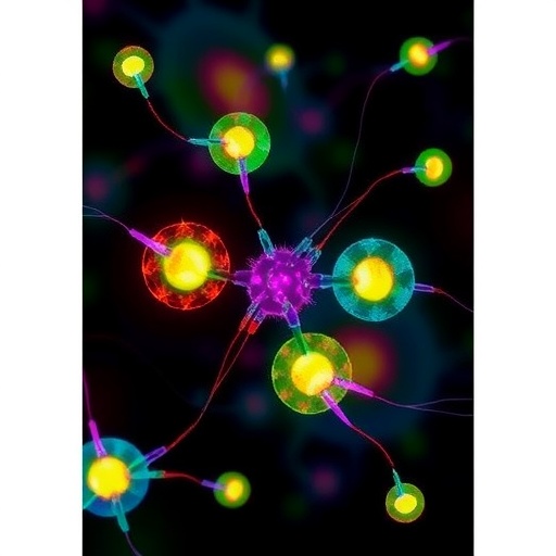

Cornell University researchers have pioneered a groundbreaking technique that transforms the way scientists observe protein behavior within living cells. By harnessing natural cellular components as intrinsic sensors, this innovative method bypasses the traditional reliance on synthetic tags, offering an unprecedented window into the dynamic inner workings of complex biological systems. This advancement holds significant promise for illuminating molecular interactions fundamental to health and disease, including viral assembly and protein misfolding implicated in cancer and neurodegeneration.
Conventional studies of protein interactions within cells often depend on invasive chemical modifications or fluorescent tags, which can disrupt native biological processes and introduce artifacts. The Cornell team circumvented these limitations by exploiting a class of naturally occurring proteins known as flavoproteins. Flavoproteins inherently contain flavins—a vitamin B2 derivative with unique magnetic properties—that act as native spin probes, enabling direct interrogation of protein environments through electron spin resonance (ESR) spectroscopy.
ESR spectroscopy, akin to magnetic resonance imaging but capable of detecting magnetic spin states at the nanoscale, traditionally requires external labeling or purified samples for accurate measurements. The Cornell researchers’ insight was to activate the magnetic spin properties of flavins inside living cells using controlled light exposure. This innovative photoactivation triggers stable magnetic spin-states detectable by ESR, rendering flavoproteins effective intrinsic reporters of molecular proximity and conformation without disrupting cellular physiology.
.adsslot_bNVwmY5RWx{width:728px !important;height:90px !important;}
@media(max-width:1199px){ .adsslot_bNVwmY5RWx{width:468px !important;height:60px !important;}
}
@media(max-width:767px){ .adsslot_bNVwmY5RWx{width:320px !important;height:50px !important;}
}
ADVERTISEMENT
The implications of this technology are profound. It enables researchers to study protein complex assembly, intermolecular interactions, and conformational changes under truly native conditions. For example, the team applied this method to investigate Aer, a membrane-bound receptor in Escherichia coli bacteria that senses oxygen. Previous studies of Aer’s assembly were restricted to artificially reconstituted systems, but this approach allowed the direct observation of Aer receptor organization within live bacterial cells for the first time.
Through ESR measurements, the researchers determined the precise distance between flavins within Aer dimers with angstrom-level accuracy. Their findings confirmed the presence of dimeric structures and unveiled the formation of higher-order arrays—large molecular assemblies that amplify signaling and are inherently unstable outside the cellular membrane environment. This represents a major breakthrough in understanding how bacterial cells spatially organize sensory proteins to respond to environmental cues.
Beyond detecting naturally occurring flavoproteins, the team engineered a novel flavoprotein variant called iLOV. This small, genetically encodable probe can be fused to virtually any protein of interest, effectively serving as a built-in molecular tag visible to ESR in living cells. The versatility of iLOV expands the potential applications of this technology across diverse biological systems, enabling the study of protein localization and interactions with unprecedented resolution and minimal perturbation.
This research challenges the previous notion that ESR spectroscopy is confined to purified molecules in vitro. By demonstrating that ESR can be employed directly in living systems, the Cornell group opens new avenues to explore the structural dynamics of proteins in their native cellular context, bridging a critical gap between biophysical techniques and cell biology. The ability to monitor proteins with such exquisite detail inside live cells represents a transformative tool for biomedical research.
Central to this approach is the advantage of using the cell’s endogenous flavoproteins as durable, non-invasive sensors of molecular interactions. The method leverages the inherent stability of flavin magnetic spin-states in cellular environments, a property previously underestimated. By activating these states with precise light stimulation, researchers can generate clear ESR signals without introducing synthetic spin labels that often compromise cell viability or function.
The strategic emphasis on vibrantly studying protein assemblies implicated in disease also marks this advance as highly relevant for understanding pathological mechanisms. For example, many neurodegenerative diseases and cancers arise from protein misfolding and aggregation, processes notoriously difficult to dissect in situ. This native ESR sensing approach could elucidate these events with high spatial and temporal resolution, providing valuable insights for therapeutic strategies.
The interdisciplinary collaboration that led to this discovery draws on expertise in chemistry, molecular biology, and advanced spectroscopy, highlighting the integrative nature of modern scientific innovation. Contributions from researchers such as Timothée Chauviré and Jack H. Freed helped refine the method’s technical robustness and applicability. Their efforts underscore the importance of multidisciplinary teams in overcoming the challenges of studying complex living systems.
Looking ahead, the Cornell team is working to adapt this novel ESR technique for use in mammalian cells and more intricate organisms. Expanding the method’s applicability to higher eukaryotes could enable tracking of protein dynamics during processes like infection, immune response, and cellular differentiation in real time. The prospect of non-invasively decoding molecular interactions in diverse biological contexts may revolutionize both fundamental research and clinical diagnostics.
This pioneering work was supported by funding from prestigious agencies including the U.S. National Science Foundation and the National Institutes of Health, underscoring its scientific merit and potential impact. The study’s publication in Nature Communications not only validates its significance but ensures broad dissemination within the scientific community, fueling future discoveries inspired by this innovative paradigm.
By demonstrating that the cell’s own machinery can serve as a sensitive, genetically encoded platform for ESR spectroscopy, Cornell researchers have introduced a powerful new lens to peer inside live cells. This approach promises to advance our grasp of molecular mechanisms underpinning life and disease, signaling a new era for the visualization of protein landscapes at the nanoscale — all without disturbing the delicate cellular balance.
Subject of Research: In-cell electron spin resonance (ESR) spectroscopy utilizing native and engineered flavoproteins as intrinsic spin probes to study protein structure and interactions inside living cells.
Article Title: Flavoproteins as native and genetically encoded spin probes for in cell ESR spectroscopy
News Publication Date: July 1, 2025
Web References: https://www.nature.com/articles/s41467-025-60623-6
Keywords
Cell biology, protein interactions, electron spin resonance, flavoproteins, live-cell spectroscopy, molecular assembly, protein structure, viral assembly, neurodegeneration, cancer biology, genetic encoding, advanced imaging technologies
Tags: advanced imaging techniques in biologycancer and neurodegeneration protein studiesCornell University research breakthroughselectron spin resonance spectroscopy applicationsflavoproteins as biological probesimplications for health and disease researchinnovative methods in molecular biologyintrinsic sensors for protein interactionsnatural sensors in cellular biologynon-invasive protein interaction studiesphotoactivation of flavins in cellsprotein behavior observation techniques


