Imagine owning a camera so powerful it can take freeze-frame photographs of a moving electron – an object traveling so fast it could circle the Earth many times in a matter of a second. Researchers at the University of Arizona have developed the world’s fastest electron microscope that can do just that.
Imagine owning a camera so powerful it can take freeze-frame photographs of a moving electron – an object traveling so fast it could circle the Earth many times in a matter of a second. Researchers at the University of Arizona have developed the world’s fastest electron microscope that can do just that.
They believe their work will lead to groundbreaking advancements in physics, chemistry, bioengineering, materials sciences and more.
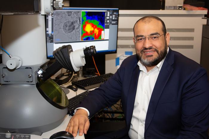
“When you get the latest version of a smartphone, it comes with a better camera,” said Mohammed Hassan, associate professor of physics and optical sciences. “This transmission electron microscope is like a very powerful camera in the latest version of smart phones; it allows us to take pictures of things we were not able to see before – like electrons. With this microscope, we hope the scientific community can understand the quantum physics behind how an electron behaves and how an electron moves.”
Hassan led a team of researchers in the departments of physics and optical sciences that published the research article “Attosecond electron microscopy and diffraction” in the Science Advances journal. Hassan worked alongside Nikolay Golubev, assistant professor of physics; Dandan Hui, co-lead author and former research associate in optics and physics who now works at the Xi’an Institute of Optics and Precision Mechanics, Chinese Academy of Sciences; Husain Alqattan, co-lead author, U of A alumnus and assistant professor of physics at Kuwait University; and Mohamed Sennary, a graduate student studying optics and physics.
A transmission electron microscope is a tool used by scientists and researchers to magnify objects up to millions of times their actual size in order to see details too small for a traditional light microscope to detect. Instead of using visible light, a transmission electron microscope directs beams of electrons through whatever sample is being studied. The interaction between the electrons and the sample is captured by lenses and detected by a camera sensor in order to generate detailed images of the sample.
Ultrafast electron microscopes using these principles were first developed in the 2000’s and use a laser to generate pulsed beams of electrons. This technique greatly increases a microscope’s temporal resolution – its ability to measure and observe changes in a sample over time. In these ultrafast microscopes, instead of relying on the speed of a camera’s shutter to dictate image quality, the resolution of a transmission electron microscope is determined by the duration of electron pulses.
The faster the pulse, the better the image.
Ultrafast electron microscopes previously operated by emitting a train of electron pulses at speeds of a few attoseconds. An attosecond is one quintillionth of a second. Pulses at these speeds create a series of images, like frames in a movie – but scientists were still missing the reactions and changes in an electron that takes place in between those frames as it evolves in real time. In order to see an electron frozen in place, U of A researchers, for the first time, generated a single attosecond electron pulse, which is as fast as electrons moves, thereby enhancing the microscope’s temporal resolution, like a high-speed camera capturing movements that would otherwise be invisible.
Hassan and his colleagues based their work on the Nobel Prize-winning accomplishments of Pierre Agostini, Ferenc Krausz and Anne L’Huilliere, who won the Novel Prize in Physics in 2023 after generating the first extreme ultraviolet radiation pulse so short it could be measured in attoseconds.
Using that work as a steppingstone, U of A researchers developed a microscope in which a powerful laser is split and converted into two parts – a very fast electron pulse and two ultra-short light pulses. The first light pulse, known as the pump pulse, feeds energy into a sample and causes electrons to move or undergo other rapid changes. The second light pulse, also called the “optical gating pulse” acts like a gate by creating a brief window of time in which the gated, single attosecond electron pulse is generated. The speed of the gating pulse therefore dictates the resolution of the image. By carefully synchronizing the two pulses, researchers control when the electron pulses probe the sample to observe ultrafast processes at the atomic level.
“The improvement of the temporal resolution inside of electron microscopes has been long anticipated and the focus of many research groups – because we all want to see the electron motion,” Hassan said. “These movements happen in attoseconds. But now, for the first time, we are able to attain attosecond temporal resolution with our electron transmission microscope – and we coined it ‘attomicroscopy.’ For the first time, we can see pieces of the electron in motion.”
Journal
Science Advances
DOI
10.1126/sciadv.adp5805
Method of Research
Computational simulation/modeling
Subject of Research
Not applicable
Article Title
Attosecond electron microscopy and diffraction
Article Publication Date
21-Aug-2024


