University of Virginia School of Medicine researchers have created an “atlas of atherosclerosis” that reveals, at the level of individual cells, critical processes responsible for forming the harmful plaque buildup that causes heart attacks, strokes and coronary artery disease.

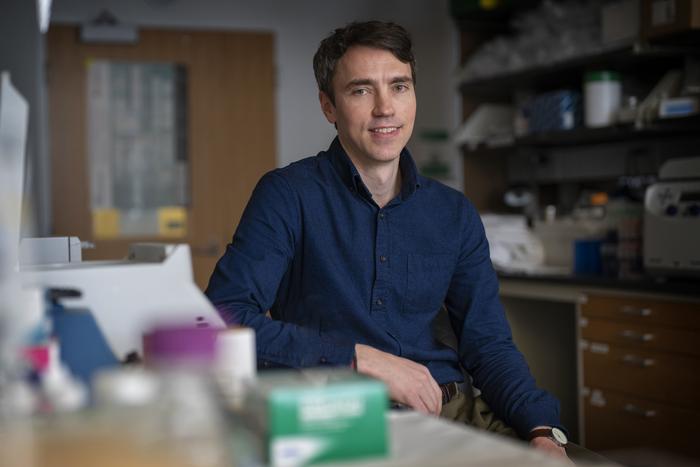
Credit: Dan Addison | University Communications
University of Virginia School of Medicine researchers have created an “atlas of atherosclerosis” that reveals, at the level of individual cells, critical processes responsible for forming the harmful plaque buildup that causes heart attacks, strokes and coronary artery disease.
Atherosclerosis, or hardening of the arteries, affects half of Americans between ages 45 and 84, and many don’t even know it, the National Institutes of Health reports. Over time, fatty plaques build up inside the arteries, where they can slow blood flow. When they break loose, they can be deadly, triggering strokes and heart attacks.
Doctors and scientists have been eager to better understand the complex factors that influence the formation and stability of the plaques, and UVA’s new work offers unprecedented insights that will facilitate the development of new ways to treat atherosclerosis, battle coronary artery disease (CAD) and help prevent plaque formation.
“To begin to develop effective treatments targeting specific disease processes in the vessel wall, we need to characterize gene expression programs at single-cell resolution,” said researcher Clint L. Miller, PhD, of the University of Virginia School of Medicine’s Center for Public Health Genomics, as well as its Departments of Biochemistry and Molecular Genetics and Public Health Sciences. “By establishing this map, we can inform strategies to reprogram dysregulated cell states in order to prevent or reverse the disease or identify biomarkers to assess a patient’s risk of having clinical events.”
Understanding Atherosclerosis
The formation of atherosclerotic plaques involves multiple types of cells, including immune cells, smooth muscle cells and endothelial cells that line the arteries. Many of these cells transition into other types of cells during plaque formation, making it a huge challenge for scientists to determine the composition and origin of the plaque itself.
Miller and his collaborators, led by graduate student Jose Verdezoto Mosquera, have built a “comprehensive single cell map of human atherosclerosis” encompassing almost 120,000 cells from atherosclerotic coronary and carotid arteries. In addition to charting broad cell lineages, the researchers leveraged this resource to dissect more granular and rare cell subtypes within atherosclerotic plaques.
The study also reveals new insights on the changes smooth muscle cells go through during disease progression, some of which contribute to the “calcification,” or hardening, of the coronary arteries. This led to the finding that two genes, LTBP1 and CRTAC1, can serve as measures for the progression of atherosclerosis.
“Beyond characterizing cell diversity, integrating this newly built atherosclerosis single-cell reference with large-scale human genetic data was critical to start identifying disease-causing cell types and subtypes,” Mosquera said. “For example, we identified the contribution of smooth muscle cell subtypes, such as fibroblast-like and lipid-rich smooth muscle cells, as well as the genes associated with these phenotypes.”
The UVA researchers say their new atlas represents a “critical step” toward developing better, more targeted interventions to battle atherosclerosis and CAD, as well as identify candidate biomarkers to prevent heart attacks and strokes and improve patient outcomes.
“We plan to extend this single-cell atlas with future iterations to include additional datasets from defined disease stages and patients from diverse backgrounds,” Miller said. “By integrating the corpus of single-cell data generated in the scientific community, we can mitigate sampling bias and establish more robust candidate disease mechanisms and potential interventions.”
Findings Published
The researchers have published their findings in the scientific journal Cell Reports. The research team consisted of Mosquera, Gaëlle Auguste, Doris Wong, Adam W. Turner, Chani J. Hodonsky, Catalina Alvarez Yela, Yipei Song, Qi Cheng, Christian L. Lino Cardenas, Konstantinos Theofilatos, Maxime Bos, Maryam Kavousi, Patricia A. Peyser, Manuel Mayr, Jason C. Kovacic, Johan L.M. Björkegren, Rajeev Malhotra, P. Todd Stukenberg, Aloke V. Finn, Sander W. van der Laan, Chongzhi Zang, Nathan C. Sheffield and Miller. Miller has received funding from biopharmaceutical company AstraZeneca for an unrelated project; a full list of the authors’ disclosures is included in the paper.
The research was supported by the National Institutes of Health, grants R01HL148239, R01HL164577, R35GM133712, R01HL142809, R01HL159514, K01HL164687, R01HL125863, R01HL130423, R01HL135093, R01HL148167-01A1 and R01 HL141425; the American Heart Association, grants 20POST35120545, JAHA909150 and A14SFRN20840000; the Swedish Research Council and Heart Lung Foundation, grants 2018-02529 and 20170265; Fondation Leducq, “PlaqOmics” grant 18CVD02; the European Union H2020 TO_AITION grant 848146; and the Single-Cell Data Insights award from the Chan Zuckerberg Initiative LLC and Silicon Valley Community Foundation.
To keep up with the latest medical research news from UVA, subscribe to the Making of Medicine blog at http://makingofmedicine.virginia.edu.
DOI
10.1016/j.celrep.2023.113380


