EMBARGOED: Not for Release Until 01 November 2023 at 16:00 (London time)

Credit: University of Southampton
EMBARGOED: Not for Release Until 01 November 2023 at 16:00 (London time)
The bodies of starfish and other echinoderms are more like heads, according to new research involving the University of Southampton.
The research, published today [1 November] in Nature, helps to answer the mystery of how these creatures evolved their distinctive star-shaped body, which has long puzzled scientists.
Echinoderms are a group of animals that includes starfish (or sea stars), sea urchins, and sand dollars. They have a unique ‘fivefold symmetric’ body plan, which means that their body parts are arranged in five equal sections. This is very different from their bilateral ancestors, which have a left- and right-hand side which mirror one another, as in humans and many other animals.
“How the different body parts of the echinoderms relate to those we see in other animal groups has been a mystery to scientists for as long as we’ve been studying them,” says Dr Jeff Thompson, a co-author on the study from the University of Southampton. “In their bilateral relatives, the body is divided into a head, trunk, and tail. But just looking at a starfish, it’s impossible to see how these sections relate to the bodies of bilateral animals.”
In the new study, led by Laurent Formery and Professor Chris Lowe at Stanford University, scientists compared the molecular markers of a sea star to other deuterostomes – a wider animal group that includes echinoderms and bilateral animals, like vertebrates. They share a common ancestor, so by comparing their development, the scientists could learn more about how echinoderms evolved their unique body plan.
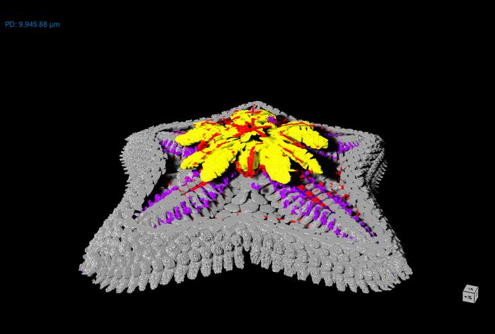
Researchers used a variety of high-tech molecular and genomic techniques to understand where different genes were expressed during the development and growth of sea stars. The team at Southampton used micro-CT scanning to understand the shape and structure of the animal in unprecedented detail.
Then, researchers at Stanford, in collaboration with Professor Dan Rokhsar at UC Berkeley and Pacific BioSciences, used ‘RNA tomography’ and ‘in situ hybridization’ to create a three-dimensional map of gene expression in the sea star and find out where specific genes are being expressed during development. Specifically, they mapped the expression of genes which control the development of the ectoderm, which includes the nervous system and the skin. This is known to mark the anterior-posterior (front-to-back) patterning in the bodies of other deuterostomes.
They found this patterning was correlated with the midline-to-lateral axis of the sea star arms – with the midline of the arm representing the front and the outmost lateral parts more like the back. In deuterostomes, there is a distinct set of genes expressed in the ectoderm of the trunk. But in the sea star, many of these genes are not expressed in the ectoderm at all.
Dr Thompson explains: “When we compared the expression of genes in a starfish to other groups of animals, like vertebrates, it appeared that a crucial part of the body plan was missing. The genes that are typically involved in the patterning of the trunk of the animal weren’t expressed in the ectoderm. It seems the whole echinoderm body plan is roughly equivalent to the head in other groups of animals.”
This suggests that sea stars and other echinoderms may have evolved their five-section body plan by losing the trunk region of their bilateral ancestors. This would have allowed the echinoderms to move and feed differently than bilaterally symmetrical animals.
“Our research tells us the echinoderm body plan evolved in a more complex way than previously thought and there is still much to learn about these intriguing creatures,” says Dr Thompson. “As someone who has studied them for the last ten years, these findings have radically changed how I think about this group of animals.”
Molecular evidence of anteroposterior patterning in adult echinoderms is published in Nature and is available online.
This research was supported by the Leverhulme Trust, NASA, NSF and the Chan Zuckerberg BioHub.
Ends
Contact
Steve Williams, Media Relations, University of Southampton [email protected] or 023 8059 3212.
Notes for editors
- Molecular evidence of anteroposterior patterning in adult echinoderms is published in Nature. An advanced copy of the paper is available upon request.
- For Interviews with Dr Jeff Thompson please contact Steve Williams, Media Relations, University of Southampton [email protected] or 023 8059 3212.
- Video and various stills available here: https://safesend.soton.ac.uk/pickup?claimID=mxmvqtmoa28Hv84n&claimPasscode=ejTaKz87vmBP4NNu
Caption: Micro-CT scan of sea star showing the skeleton (grey), digestive system (yellow), nervous system (blue), muscles (red) and water vascular system (purple). Credit: University of Southampton
Additional information
The University of Southampton drives original thinking, turns knowledge into action and impact, and creates solutions to the world’s challenges. We are among the top 100 institutions globally (QS World University Rankings 2023). Our academics are leaders in their fields, forging links with high-profile international businesses and organisations, and inspiring a 22,000-strong community of exceptional students, from over 135 countries worldwide. Through our high-quality education, the University helps students on a journey of discovery to realise their potential and join our global network of over 200,000 alumni. www.southampton.ac.uk
www.southampton.ac.uk/news/contact-press-team.page
Follow us on X: https://twitter.com/UoSMedia
Journal
Nature
DOI
10.1038/s41586-023-06669-2
Article Title
‘Molecular evidence of anteroposterior patterning in adult echinoderms
Article Publication Date
1-Nov-2023


