Reston, VA—A novel nuclear medicine imaging protocol can take the place of two separate imaging scans for the evaluation of brain changes linked to cognitive impairment, cutting costs and radiation exposure for patients as well as providing them greater comfort. The protocol—known as dual-phase amyloid PET—can assess both amyloid deposition and neurodegeneration with a single tracer injection, assisting physicians as they classify patients on the Alzheimer’s disease continuum. This research was published in the February issue of The Journal of Nuclear Medicine.
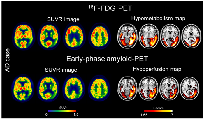
Credit: Image created by C Boccalini, et al., University of Geneva, Geneva, Switzerland.
Reston, VA—A novel nuclear medicine imaging protocol can take the place of two separate imaging scans for the evaluation of brain changes linked to cognitive impairment, cutting costs and radiation exposure for patients as well as providing them greater comfort. The protocol—known as dual-phase amyloid PET—can assess both amyloid deposition and neurodegeneration with a single tracer injection, assisting physicians as they classify patients on the Alzheimer’s disease continuum. This research was published in the February issue of The Journal of Nuclear Medicine.
Alzheimer’s disease causes several changes in the brain, including neurodegeneration and beta-amyloid deposition. Currently, 18F-FDG scans are used to measure brain metabolism, which can indicate neurodegeneration. Amyloid PET scans are performed to evaluate beta-amyloid deposition, a key characteristic of Alzheimer’s disease.
“During the amyloid PET scans, nuclear medicine physicians wait at least 50 minutes after the radiotracer injection to acquire the image of beta-amyloid deposition. If images are also taken immediately after injection (early-phase), they can provide a measure of brain perfusion. Since brain perfusion and brain metabolism are closely related, the perfusion information could replace an 18F-FDG PET scan,” said Cecilia Boccalini, MSc, PhD candidate at the Laboratory of Neuroimaging and Innovative Molecular Tracers and faculty of medicine at the University of Geneva Neurocenter in Geneva, Switzerland and faculty of neuroscience at Vita-Salute San Raffaele University in Milan, Italy.
The study aimed to compare early-phase amyloid PET scans and 18F-FDG scans at the individual level, as well as their ability to distinguish patients along the Alzheimer’s disease continuum. A total of 166 patients ranging from cognitively unimpaired to mild cognitive impairment and dementia underwent 18F-FDG and early-phase amyloid PET scans. Brain hypoperfusion maps (from the early-phase amyloid PET) and brain hypometabolism maps (from 18F-FDG PET) were created and compared for each patient.
Results showed that brain hypoperfusion assessed by early-phase amyloid PET is comparable with brain hypometabolism assessed by 18F-FDG-PET. Hypoperfusion and hypometabolism patterns were also equally able to distinguish patients with neurodegenerative diseases from controls.
“This dual-phase amyloid-PET protocol allows physicians to measure brain amyloid and perfusion in a single procedure,” stated Valentina Garibotto, MD, head of the Nuclear Medicine and Molecular Imaging Division at Geneva University Hospitals in Geneva, Switzerland, and associate professor at the University of Geneva. “Obtaining both amyloid and perfusion data with a single tracer injection is optimal in terms of patient safety and radiation exposure, comfort, and costs. Our work supports the routine use of the dual-phase amyloid PET protocol in clinical practice.”
This study was made available online in July 2022.
The authors of “Early-phase 18F-Florbetapir and 18F-Flutemetamol images as proxies of brain metabolism in a memory clinic setting” include Cecilia Boccalini, Laboratory of Neuroimaging and Innovative Molecular Tracers (NIMTlab), Geneva University Neurocenter and Faculty of Medicine, University of Geneva, Switzerland, Vita-Salute San Raffaele University, Milan, Italy, and In Vivo Human Molecular and Structural Neuroimaging Unit, Division of Neuroscience, IRCCS San Raffaele Scientific Institute, Milan, Italy; Débora Elisa Peretti and Sara Stampacchia, Laboratory of Neuroimaging and Innovative Molecular Tracers (NIMTlab), Geneva University Neurocenter and Faculty of Medicine, University of Geneva, Switzerland; Federica Ribaldi and Giovanni B. Frisoni, Laboratory of Neuroimaging of Aging (LANVIE), University of Geneva, Geneva, Switzerland and Memory Clinic, Geneva University Hospitals, Geneva, Switzerland; Max Scheffler, Division of Radiology, Diagnostic Department, Geneva University Hospitals, Geneva, Switzerland; Szymon Tomczyk, Laboratory of Neuroimaging of Aging (LANVIE), University of Geneva, Geneva, Switzerland; Cristelle Rodriguez and Panteleimon Giannakopoulos, Division of Institutional Measures, Medical Direction, University Hospitals of Geneva, Geneva, Switzerland and Department of Psychiatry, Faculty of Medicine, University of Geneva, Geneva, Switzerland; Marie-Louise Montandon, Department of Psychiatry, Faculty of Medicine, University of Geneva, Geneva, Switzerland and Department of Rehabilitation and Geriatrics, Geneva University Hospitals and University of Geneva, Geneva, Switzerland; Sven Haller, CIMC – Centre d’Imagerie Médicale de Cornavin, Geneva, Switzerland, Faculty of Medicine of the University of Geneva, Geneva, Switzerland, Department of Surgical Sciences, Radiology, Uppsala University, Uppsala, Sweden, and Department of Radiology, Beijing Tiantan Hospital, Capital Medical University, Beijing, China; Daniela Perani, Vita-Salute San Raffaele University, Milan, Italy, In Vivo Human Molecular and Structural Neuroimaging Unit, Division of Neuroscience, IRCCS San Raffaele Scientific Institute, Milan, Italy, and Nuclear Medicine Unit, San Raffaele Hospital, Milan, Italy; and Valentina Garibotto, Laboratory of Neuroimaging and Innovative Molecular Tracers (NIMTlab), Geneva University Neurocenter and Faculty of Medicine, University of Geneva, Switzerland, Division of Nuclear Medicine and Molecular Imaging, Geneva University Hospitals, Geneva, Switzerland, and CIBM Center for Biomedical Imaging, Geneva, Switzerland.
Visit the JNM website for the latest research, and follow our new Twitter and Facebook pages @JournalofNucMed or follow us on LinkedIn.
###
Please visit the SNMMI Media Center for more information about molecular imaging and precision imaging. To schedule an interview with the researchers, please contact Rebecca Maxey at (703) 652-6772 or [email protected].
About JNM and the Society of Nuclear Medicine and Molecular Imaging
The Journal of Nuclear Medicine (JNM) is the world’s leading nuclear medicine, molecular imaging and theranostics journal, accessed 15 million times each year by practitioners around the globe, providing them with the information they need to advance this rapidly expanding field. Current and past issues of The Journal of Nuclear Medicine can be found online at http://jnm.snmjournals.org.
JNM is published by the Society of Nuclear Medicine and Molecular Imaging (SNMMI), an international scientific and medical organization dedicated to advancing nuclear medicine and molecular imaging—precision medicine that allows diagnosis and treatment to be tailored to individual patients in order to achieve the best possible outcomes. For more information, visit www.snmmi.org.
Journal
Journal of Nuclear Medicine
DOI
10.2967/jnumed.122.264256
Article Title
Early-phase 18F-Florbetapir and 18F-Flutemetamol images as proxies of brain metabolism in a memory clinic setting
Article Publication Date
1-Feb-2023
COI Statement
Cecilia Boccalini was supported by an IBRO Exchange Fellowship.


