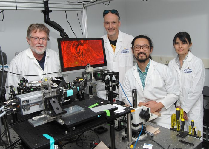
WASHINGTON — Researchers have shown that a new microscopy technique can capture dynamic 3D images of an entire zebrafish larvae while maintaining cellular resolution in all three dimensions. By giving scientists an unprecedented view of how cells interact in their most natural state, the new technique could provide information that is used to develop new treatments for diseases, for example.
Credit: Photo by Mike McElwaine
WASHINGTON — Researchers have shown that a new microscopy technique can capture dynamic 3D images of an entire zebrafish larvae while maintaining cellular resolution in all three dimensions. By giving scientists an unprecedented view of how cells interact in their most natural state, the new technique could provide information that is used to develop new treatments for diseases, for example.
“Unraveling underlying cellular structures and their interaction is fundamentally important to understanding life,” said research team leader Ji Yi of Johns Hopkins University. “However, the limitations of light diffraction make it difficult to image with 3D cellular resolution over large areas of several millimeters. We circumvent the trade-offs between field of view, depth resolution and imaging speed to achieve 4D cellular resolution over a much larger field of view than previously possible.”
In Optica, the Optica Publishing Group’s journal for high-impact research, the researchers report that their new mesoscopic oblique plane microscopy method can capture up to three times more resolvable image points within an imaged 3D volume compared to other similar systems. Oblique plane microscopy is a type of light-sheet microscopy, which uses a sheet of laser light to illuminate a thin slice of a sample labeled with fluorescent markers.
“Looking at biological systems in their larger context — also known as a mesoscopic scale — provides holistic information for complex biological systems such as a complete neural circuit in mouse brain, 3D tissue culture or entire zebrafish larvae,” said Yi. “Our work also lays a foundation for further development that would allow even faster, larger and deeper biological imaging.”
Overcoming trade-offs
Whether imaging with a microscope or a smartphone camera, it is difficult to use a single set of optics to acquire both large scene coverage and enough resolution to see details. While modern smartphones often use more than one camera set to overcome this challenge, this isn’t typically feasible with microscopy.
“Instead of adding another camera set, we used an optical component known as a transmission grating to create a diffractive light sheet,” said Yi. “This improves depth sectioning and resolution while using a low magnification lens. The result is the ability to perform mesoscopic scale imaging over a field of view that is several millimeters wide while still being able to resolve individual cells in 3D.”
The researchers demonstrated their new technique by using it to image two important large-scale model systems: living zebrafish larvae that were 3-4 mm long and mouse brain slices in which the cells are kept alive. Both expressed fluorescence proteins that labeled specific cell types such as neurons and blood cells.
Using their mesoscopic oblique plane microscopy technique, the researchers were able to image a field of view measuring up to 5.4 × 3.3 millimeters with a resolution of 2.5× 3 × 6 microns, which allowed volumetric imaging of 3D cellular structures with a single scan. For the zebrafish larvae, they captured whole-body volumetric recordings of neuronal activity at 2 Hz volume rate, uniquely enabling studies on neural circuits over the entire central nervous system in a vertebrate. They also showed whole-body volumetric recordings of blood flow dynamics at 5 Hz with 3D cellular resolution, to allow single-cell tracking within the complete 3D circulation system for the first time.
Moving forward, the researchers would like to improve the light collection efficiency to further increase the imaging speed. They also want to incorporate multiphoton imaging to allow better penetration depth, another long-standing challenge in optical imaging.
Paper: W. Shao, M. Chang, K. Emmerich, P. O. Kanold, J. S. Mumm, J. Yi, “Mesoscopic oblique plane microscopy (Meso-OPM) with a diffractive light sheet- enabling large-scale 4D cellular resolution imaging,” 9,12 (2022).
DOI: doi.org/10.1364/OPTICA.471101
About Optica
Optica is an open-access journal dedicated to the rapid dissemination of high-impact peer-reviewed research across the entire spectrum of optics and photonics. Published monthly by Optica Publishing Group, the Journal provides a forum for pioneering research to be swiftly accessed by the international community, whether that research is theoretical or experimental, fundamental or applied. Optica maintains a distinguished editorial board of more than 60 associate editors from around the world and is overseen by Editor-in-Chief Prem Kumar, Northwestern University, USA. For more information, visit Optica.
About Optica Publishing Group (formerly OSA)
Optica Publishing Group is a division of Optica (formerly OSA), Advancing Optics and Photonics Worldwide. It publishes the largest collection of peer-reviewed content in optics and photonics, including 18 prestigious journals, the society’s flagship member magazine, and papers from more than 835 conferences, including 6,500+ associated videos. With over 400,000 journal articles, conference papers and videos to search, discover and access, Optica Publishing Group represents the full range of research in the field from around the globe.
Journal
Optica
DOI
10.1364/OPTICA.471101
Article Title
Mesoscopic oblique plane microscopy (Meso-OPM) with a diffractive light sheet- enabling large-scale 4D cellular resolution imaging
Article Publication Date
8-Dec-2022

