Weill Cornell Medicine researchers have developed a computational method to map the architecture of human tissues in unprecedented detail. Their approach promises to accelerate studies on organ-scale cellular interactions and could enable powerful new diagnostic strategies for a wide range of diseases.
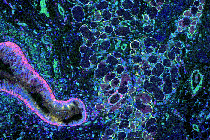
Credit: Junbum Kim
Weill Cornell Medicine researchers have developed a computational method to map the architecture of human tissues in unprecedented detail. Their approach promises to accelerate studies on organ-scale cellular interactions and could enable powerful new diagnostic strategies for a wide range of diseases.
The method, published Oct. 31 in Nature Methods, grew out of the scientists’ frustration with the gap between classical microscopy and modern single-cell molecular analysis. “Looking at tissues under the microscope, you see a bunch of cells that are grouped together spatially—you see that organization in images almost immediately,” said lead author Junbum Kim, a graduate student in physiology and biophysics at Weill Cornell Medicine. “Now, cell biologists have gained the ability to examine individual cells in tremendous detail, down to which genes each cell is expressing, so they’re focused on the cells instead of focusing on the tissue structure,” he said.
However, “it’s crucial for researchers to learn more about the details of tissue structure; fundamental changes in the relationships between cells within a tissue drive both healthy and diseased organ function,” said senior author Dr. Olivier Elemento, director of the Englander Institute for Precision Medicine and a professor of physiology and biophysics and of computational genomics in computational biomedicine at Weill Cornell Medicine.
Manually combining single cell data with maps of tissue structure is slow and tedious, though. Machine learning algorithms have shown some potential for automating the process, but they’re limited by the data used to train them. To address that, Kim and his colleagues developed an unsupervised computational strategy, using a combination of single-cell gene expression profiles and cells’ locations to define structural regions within a tissue.
Co-senior author Dr. André Rendeiro, a postdoctoral fellow at Weill Cornell Medicine during the study and currently a principal investigator at the Research Center for Molecular Medicine of the Austrian Academy of Sciences in Vienna, Austria, compares the new method to mapping a city such as New York: “One way to go about it would be to go to every intersection and count each kind of building: is it residential, is it commercial … is it a shop or restaurant?” Putting all of those data into one matrix, and the buildings’ locations into another, one could then combine the two matrices and look for patterns.
“Essentially, we could start to make a general statement about where the different neighborhoods are and where their borders are based on the abundance of, say, residential versus commercial buildings — just as anyone walking through the Upper East Side, Midtown or Downtown would do based on their observations,” said Dr. Rendeiro.
The researchers used the new method to generate detailed maps of several types of tissues, identifying and quantifying new aspects of microanatomy – the patterns that emerge at small scale when cells interact and that determine the ultimate function of tissue. Collaborating with a colleague at the University of North Carolina at Chapel Hill who studies lung disease, they also demonstrated that their technique could draw fine shades of distinction between different disease states in a tissue.
While cancer and other chronic diseases often cause major changes in tissue structure, detailed microanatomy could also help in diagnosing and treating more acute conditions. Rendeiro points to severe COVID-19 as one example, where “there are a lot of immune cells that move into the neighborhood, and there’s really dramatic change in the lung tissue.” The team is now applying their new technique to a wide range of tissues to understand how changes in tissue organization underlie its function in healthy state and dysfunction in disease.
Many Weill Cornell Medicine physicians and scientists maintain relationships and collaborate with external organizations to foster scientific innovation and provide expert guidance. The institution makes these disclosures public to ensure transparency. For this information, see profile for Dr. Elemento.
Journal
Nature Methods

