Imaging method poised to bring new insight into how the brain works in health and disease
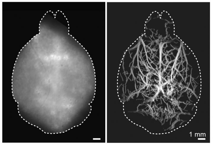
Credit: Daniel Razansky, University and ETH Zurich
WASHINGTON — Researchers have developed a new technique that allows microscopic fluorescence imaging at four times the depth limit imposed by light diffusion. Fluorescence microscopy is often used to image molecular and cellular details of the brain in animal models of various diseases but, until now, has been limited to small volumes and highly invasive procedures due to intense light scattering by the skin and skull.
“Visualization of biological dynamics in an unperturbed environment, deep in a living organism, is essential for understanding the complex biology of living organisms and progression of diseases,” said research team leader Daniel Razansky from the University of Zurich and ETH Zurich, both in Switzerland. “Our study represents the first time that 3D fluorescence microscopy has been performed fully noninvasively at capillary level resolution in an adult mouse brain, effectively covering a field of view of about 1 centimeter.”
In Optica, The Optical Society’s (OSA) journal for high impact research, the researchers describe their new technique, which is called diffuse optical localization imaging (DOLI). It takes advantage of what is known as the second near-infrared (NIR-II) spectral window from 1000 to 1700 nanometers, which exhibits less scattering.
“Enabling high-resolution optical observations in deep living tissues represents a long-standing goal in the biomedical imaging field,” said Razansky. “DOLI’s superb resolution for deep-tissue optical observations can provide functional insights into the brain, making it a promising platform for studying neural activity, microcirculation, neurovascular coupling and neurodegeneration.”
Achieving greater depth
For the new technique, the researchers intravenously inject a living mouse with fluorescent microdroplets at a concentration that creates a sparse distribution in the blood stream. Tracking these flowing targets enables reconstruction of a high-resolution map of the deep cerebral microvasculature in the mouse brain.
“The method eliminates background light scattering and is performed with the scalp and skull intact,” said Razansky. “Interestingly, we also observed strong dependence of the spot size recorded by the camera on microdroplet’s depth in the brain, which enabled depth-resolved imaging.”
The new approach benefits from the recent introduction of highly efficient short-wave infrared cameras based on InGaAs sensors. Another key building block was the use of novel contrast agents exhibiting strong fluorescence responses in the NIR-II window, such as lead sulfide (PbS)-based quantum dots.
Crisp and clear imaging
The researchers first tested the new technique in synthetic models of tissue known as tissue phantoms that mimic average brain tissue properties, demonstrating that they could acquire microscopic resolution images at depths of up to 4 millimeters in optically opaque tissues. They then performed DOLI in living mice where cerebral microvasculature as well as blood flow velocity and direction could be visualized entirely noninvasively.
The researchers are working to optimize precision in all three dimensions to improve DOLI’s resolution. They are also developing improved fluorescent agents that are smaller, have stronger fluorescence intensity and are more stable in vivo. This will significantly boost DOLI’s performance in terms of the achievable signal to noise and imaging depth.
“We expect that DOLI will emerge as a powerful approach for fluorescence imaging of living organisms at previously inaccessible depth and resolution regimes,” said Razansky. “This will greatly enhance the in vivo applicability of fluorescence microscopy and tomography techniques.”
###
Paper: Q. Zhou, Z. Chen, J. Robin, X.-L. Deán-Ben, D. Razansky, “Diffuse optical localization imaging (DOLI) enables noninvasive deep brain microangiography in NIR-II window,” Optica, 8, 6, 796-803 (2021).
DOI: https:/
About Optica
Optica is an open-access, journal dedicated to the rapid dissemination of high-impact peer-reviewed research across the entire spectrum of optics and photonics. Published monthly by The Optical Society (OSA), Optica provides a forum for pioneering research to be swiftly accessed by the international community, whether that research is theoretical or experimental, fundamental or applied. Optica maintains a distinguished editorial board of more than 60 associate editors from around the world and is overseen by Editor-in-Chief Prem Kumar, Northwestern University, USA. For more information, visit Optica.
About The Optical Society
Founded in 1916, The Optical Society (OSA) is the leading professional organization for scientists, engineers, students and business leaders who fuel discoveries, shape real-life applications and accelerate achievements in the science of light. Through world-renowned publications, meetings and membership initiatives, OSA provides quality research, inspired interactions and dedicated resources for its extensive global network of optics and photonics experts. For more information, visit osa.org.
Media Contacts:
Aaron Cohen
(301) 633-6773
[email protected]
Media Contact
James Merrick
[email protected]
Related Journal Article
http://dx.