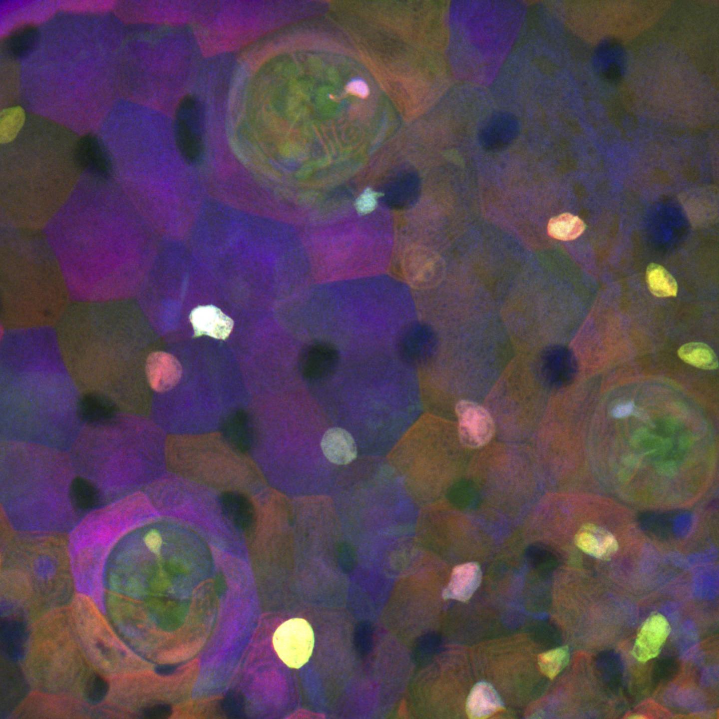
Capturing highly motile and invasive neuromast-associated ionocytes

Credit: Piotrowski Lab
KANSAS CITY, MO–One of the evolutionary disadvantages for mammals, relative to other vertebrates like fish and chickens, is the inability to regenerate sensory hair cells. The inner hair cells in our ears are responsible for transforming sound vibrations and gravitational forces into electrical signals, which we need to detect sound and maintain balance and spatial orientation. Certain insults, such as exposure to noise, antibiotics, or age, cause inner ear hair cells to die off, which leads to hearing loss and vestibular defects, a condition reported by 15% of the US adult population. In addition, the ion composition of the fluid surrounding the hair cells needs to be tightly controlled, otherwise hair cell function is compromised as observed in Ménière’s disease.
While prosthetics like cochlear implants can restore some level of hearing, it may be possible to develop medical therapies to restore hearing through the regeneration of hair cells. Investigator Tatjana Piotrowski, PhD, at the Stowers Institute for Medical Research is part of the Hearing Restoration Project of the Hearing Health Foundation, which is a consortium of laboratories that do foundational and translational science using fish, chicken, mouse, and cell culture systems.
“To gain a detailed understanding of the molecular mechanisms and genes that enable fish to regenerate hair cells, we need to understand which cells give rise to regenerating hair cells and related to that question, how many cell types exist in the sensory organs,” says Piotrowski.
The Piotrowski Lab studies regeneration of sensory hair cells in the zebrafish lateral line. Located superficially on the fish’s skin, these cells are easy to visualize and to access for experimentation. The sensory organs of the lateral line, known as neuromasts, contain support cells which can readily differentiate into new hair cells. Others had shown, using techniques to label cells of the same embryonic origin in a particular color, that cells within the neuromasts derive from ectodermal thickenings called placodes.
It turns out that while most cells of the zebrafish neuromast do originate from placodes, this isn’t true for all of them.
In a paper published online April 19, 2021, in Developmental Cell, researchers from the Piotrowski Lab describe their discovery of the occasional occurrence of a pair of cells within post-embryonic and adult neuromasts that are not labeled by lateral line markers. When using a technique called Zebrabow to track embryonic cells through development, these cells are labeled a different color than the rest of the neuromast.
“I initially thought it was an artifact of the research method,” says Julia Peloggia, a predoctoral researcher at The Graduate School of the Stowers Institute for Medical Research, co-first author of this work along with another predoctoral researcher, Daniela Münch. “Especially when we are looking just at the nuclei of cells, it’s pretty common in transgenic animal lines that the labels don’t mark all of the cells,” adds Münch.
Peloggia and Münch agreed that it was difficult to discern a pattern at first. “Although these cells have a stereotypical location in the neuromast, they’re not always there. Some neuromasts have them, some don’t, and that threw us off,” says Peloggia.
By applying an experimental method called single-cell RNA sequencing to cells isolated by fluorescence-activated cell sorting, the researchers identified these cells as ionocytes–a specialized type of cell that can regulate the ionic composition of nearby fluid¬. Using lineage tracing, they determined that the ionocytes derived from skin cells surrounding the neuromast. They named these cells neuromast-associated ionocytes.
Next, they sought to capture the phenomenon using time-lapse and high-resolution live imaging of young larvae.
“In the beginning, we didn’t have a way to trigger invasion by these cells. We were imaging whenever the microscope was available, taking as many time-lapses as possible–over days or weekends–and hoping that we would see the cells invading the neuromasts just by chance,” says Münch.
Ultimately, the researchers observed that the ionocyte progenitor cells migrated into neuromasts as pairs of cells, rearranging between other support cells and hair cells while remaining associated as a pair. They found that this phenomenon occurred all throughout early larval, later larval, and well into the adult stages in zebrafish. The frequency of neuromast-associated ionocytes correlated with developmental stages, including transfers when larvae were moved from ion-rich embryo medium to ion-poor water.
From each pair, they determined that only one cell was labeled by a Notch pathway reporter tagged with fluorescent red or green protein. To visualize the morphology of both cells, they used serial block face scanning electron microscopy to generate high-resolution three-dimensional images. They found that both cells had extensions reaching the apical or top surface of the neuromast, and both often contained thin projections. The Notch-negative cell displayed unique “toothbrush-like” microvilli projecting into the neuromast lumen or interior, reminiscent of that seen in gill and skin ionocytes.
“Once we were able to see the morphology of these cells–how they were really protrusive and interacting with other cells–we realized they might have a complex function in the neuromast,” says Münch.
“Our studies are the first to show that ionocytes invade sensory organs even in adult animals and that they only do so in response to changes in the environment that the animal lives in,” says Peloggia. “These cells therefore likely play an important role allowing the animal to adapt to changing environmental conditions.”
Ionocytes are known to exist in other organ systems. “The inner ear of mammals also contains cells that regulate the ion composition of the fluid that surrounds the hair cells, and dysregulation of this equilibrium leads to hearing and vestibular defects,” says Piotrowski. While ionocyte-like cells exist in other systems, it’s not known whether they exhibit such adaptive and invasive behavior.
“We don’t know if ear ionocytes share the same transcriptome, or collection of gene messages, but they have similar morphology to an extent and may possibly have a similar function, so we think they might be analogous cells,” says Münch. Our discovery of neuromast ionocytes will let us test this hypothesis, as well as test how ionocytes modulate hair cell function at the molecular level,” says Peloggia.
Next, the researchers will focus on two related questions–what causes these ionocytes to migrate and invade the neuromast, and what is their specific function?
“Even though we made this astounding observation that ionocytes are highly motile, we still don’t know how the invasion is triggered,” says Peloggia. “Identifying the signals that attract ionocytes and allow them to squeeze into the sensory organs might also teach us how cancer cells invade organs during disease.” While Peloggia plans to investigate what triggers the cells to differentiate, migrate, and invade, Münch will focus on characterizing the function of the neuromast-associated ionocytes. “The adaptive part is really interesting,” explains Münch. “That there is a process involving ionocytes extending into adult stages that could modulate and change the function of an organ–that’s exciting.”
Other coauthors of the study include Paloma Meneses-Giles, Andrés Romero-Carvajal, PhD, Mark E. Lush, PhD, and Melainia McClain from Stowers; Nathan D. Lawson from the University of Massachusetts Medical School; and Y. Albert Pan, PhD, from Virginia Tech Carilion.
The work was funded by the Stowers Institute for Medical Research and the National Institute of Child Health and Human Development of the National Institutes of Health (award 1R01DC015488-01A1). The content is solely the responsibility of the authors and does not necessarily represent the official views of the NIH.
Lay Summary of Findings
Humans cannot regenerate inner ear hair cells, which are responsible for detecting sound, but non-mammalian vertebrates can readily regenerate sensory hair cells that are similar in function. During the quest to understand zebrafish hair cell regeneration, researchers from the lab of Investigator Tatjana Piotrowski, PhD, at the Stowers Institute for Medical Research discovered the existence of a cell type not previously described in the process.
The research team found newly differentiated, migratory, and invasive ionocytes located in the sensory organs that house the cells giving rise to new hair cells in larval and adult fish. The researchers published their findings online April 19, 2021, in Developmental Cell. Normal invasive (that is, non-metastatic) behavior of cells after embryonic development is not often observed. Future research by the team will focus on identifying triggers for such behavior and the function of such cells, including how this process may relate to hair cell regeneration.
###
About the Stowers Institute for Medical Research
Founded in 1994 through the generosity of Jim Stowers, founder of American Century Investments, and his wife, Virginia, the Stowers Institute for Medical Research is a non-profit, biomedical research organization with a focus on foundational research. Its mission is to expand our understanding of the secrets of life and improve life’s quality through innovative approaches to the causes, treatment, and prevention of diseases.
The Institute consists of twenty independent research programs. Of the approximately 500 members, over 370 are scientific staff that includes principal investigators, technology center directors, postdoctoral scientists, graduate students, and technical support staff. Learn more about the Institute at http://www.
Media Contact
Kimberly Bland, PhD
[email protected]


