Credit: by Tonmoy Chakraborty, Bingying Chen, Stephan Daetwyler, Bo-Jui Chang, Oliver Vanderpoorten, Etai Sapoznik, Clemens Kaminski, Tuomas P.J. Knowles, Kevin M. Dean, and Reto Fiolka
Fast imaging is of great interest in microscopy, computer vision, and laser machining. For example, in neuroscience, high-speed volumetric imaging is essential to monitor dynamic biological processes, including membrane voltage activity (with dynamics on the time scale of 1 ms or less) or cerebral blood flow. How fast one can image is tightly connected to how fast one can change the position of the focus of the imaging system, particularly in the third dimension.
Traditional ways to refocus do so by either mechanically moving the microscope objective or the sample, which both leads to low scanning speed in the third dimension as the speed of moving physical objects is limited by inertia. A potential way to alleviate this problem is through remote focusing, which realizes refocusing by changing the wavefront of the optical system. However, most of the existing technologies face the tradeoff between resolution and speed. As such, there remains a need for a 3D scan technology capable of reaching multi-kHz rates while avoiding aberrations that would lower its resolution.
In a manuscript published in Light Science & Application, a team of scientists, led by Professor Reto Fiolka from the Department of Cell Biology and Lyda Hill Department of Bioinformatics, at UT Southwestern Medical Center, Dallas, TX, USA., and co-workers have developed a novel optical design to overcome these challenges. They employed well-established lateral scan technologies and transformed the lateral scan motion into refocusing in the third dimension to realize high-speed volumetric imaging. They took the concept of aberration-free remote focusing, and instead of moving a corresponding remote mirror in the third dimension, they scanned a laser spot laterally with a high-speed galvanometer over a stationary mirror. If the distance between the stationary mirror and the objective lens is not constant along the scan direction, a defocus will be introduced as is necessary for remote refocusing. Furthermore, on the return path, the lateral scan component is perfectly compensated, such that a pure scan motion in the third dimension is obtained. Thereby, the researchers were able to harness high-speed lateral scan technologies to rapidly move a high-resolution laser focus in the third dimension.
Two implementations using a step mirror and a tilted planar mirror, were adopted to realize this concept. The former allows arbitrarily large axial step sizes over a finite number of steps, and the latter allows for an arbitrary number and size of axial steps and is capable of continuous scanning in the third dimension, albeit over a more limited scan range. With the two implementations, the scientists introduce applications of this technology:
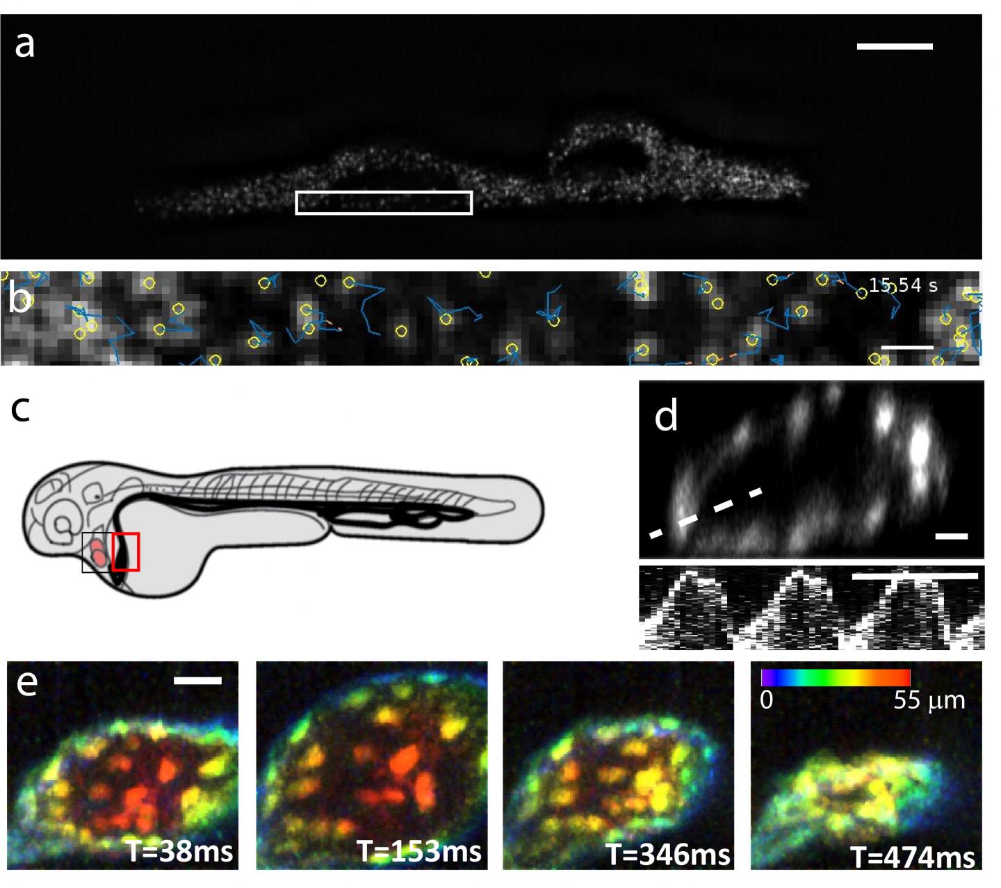
“Our first practical demonstration on microscopic imaging was accelerating axially swept light-sheet microscopy (ASLM), which has been criticized for its slow acquisition speed (around 10 Hz framerate in high-resolution implementations, previously). Our new scan technology allows one order of magnitude acceleration while keeping the high spatial resolving power of this emerging imaging technology. In a second application, we implemented our scanning technology in a 2-photon raster scanning microscope and performed high-resolution volumetric imaging with a scan rate in the third dimension of 12 kHz. Indeed, at this spatial resolution, our approach is 6-fold faster than previously reported aberration-free focusing technologies. We then demonstrated the potential of our technology for intravital microscopy by imaging the beating heart of a zebrafish embryo. We believe that this opens up major applications for intravital imaging, especially in the neurosciences.”
“Both the discrete and continuous scan technologies may find many applications to image different layers of the brain nearly simultaneously or to rapidly acquire whole volumes to measure neuronal firing patterns or cerebral blood flow. Importantly and unlike previous technologies, our approach is fully compatible with acousto-optical deflectors and thus theoretically capable of scanning on the sub-microsecond timescale (e.g., > 1 MHz) in the third dimension. Thus, using resonant Lissajous scanning patterns, we foresee the possibility for volumetric imaging at kHz rates.” the scientists forecast.
###
Media Contact
Reto Fiolka
[email protected]
Related Journal Article
http://dx.


