UNIGE scientists have demonstrated that cellular tissues are deformed by buckling (or bending under the action of compression), a phenomenon that could lie behind embryo morphogenesis.
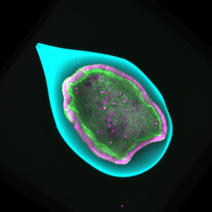
Credit: @UNIGE/Aurélien Roux
The embryo of an animal first looks like a hollow sphere. Invaginations then appear at different stages of development, which will give rise to the body’s structures (the brain, digestive tract, etc.). According to a hypothesis that dates back more than a century, buckling could be the dominant mechanism that triggers invagination – buckling being a term that describes the lateral deformation of a material under compression. Although this explanation has long won the support of biologists, it has never been subjected to formal proof, mainly because of the difficulty – if not the impossibility – of measuring the tiny forces involved. This gap has finally been filled thanks to a study carried out by a multidisciplinary team of scientists from the University of Geneva (UNIGE). This tour de force, published in the journal Developmental Cell, owes its success to a long collaboration between specialists in biological experimentation, analytical theoretical physics and computer simulation.
“The basic question underpinning our work is to find out how to shape cellular tissue,” begins Aurélien Roux, a professor in the Department of Biochemistry in UNIGE’s Faculty of Science. Observing embryo development has made it possible to describe several mechanisms that are at work. One of these is apical constriction: a local curvature of the surface of the embryo under the effect of a coordinated deformation of the cells themselves (their “apex” tightens and their “base” relaxes). But, as Professor Roux continues: “This mechanism is by no means powerful enough to explain the appearance of major invaginations during the development of the blastocyst (one of the early stages of the embryo).”
A century ago, biologists suggested that buckling is the physical mechanism that generates these deep folds. The same phenomenon is observed when you flatten a sheet of paper and bring the two opposite edges together: the middle of the sheet rises. In the case of embryos, the lateral force comes from cells which, when they proliferate, exert increasing pressure on the surface. Moreover, this surface is confined in a vitelline envelope, which – although it is elastic – prevents any spatial expansion.
Since the description of the phenomenon is quite eloquent and the analogies in nature are legion, the explanation easily won consensus in the biologist community. It has long been unthinkable to measure the forces present on the surface of embryos in order to verify that it really is a question of buckling (which obeys the well-known laws of material physics) and not another mechanism.
Analytical, IT and biological approaches
Nevertheless, the Geneva scientists – keen to provide quantitative proof of the phenomenon – conducted a long-term study. Anastasiya Trushko, a researcher the Department of Biochemistry, and Professor Roux managed to manufacture small envelopes with all the physical properties of the natural vitelline. They also succeeded in growing a monolayer formed of a hundred cells on the inner surface. These small models, less than half a millimetre in diameter, were perfectly controlled under laboratory conditions, and were used to recreate the phenomenon of invagination in vitro and to study it under microscope. The forces involved were determined in particular thanks to small variations in the thickness of the envelope of the artificial embryos.
Meanwhile Carles Blanch-Mercader and Karsten Kruse, respectively a researcher and professor in UNIGE’s Departments of Biochemistry and Theoretical Physics, used the measurements to show that the relationship between the strength and shape of the artificial embryos was as expected for buckling. With the help of material physics equations, they were able to extract the macroscopic mechanical parameters from the cellular tissues, such as their stiffness.
Finally, in order to link these macroscopic characteristics to biological processes at cellular level, Aziza Merzouki and Bastien Chopard – respectively a researcher and professor in the Computer Science Department at UNIGE – simulated the development of the embryo by computer, viewing it as a set of independent cells. “The IT approach gives the unique possibility of observing certain aspects of the phenomenon that are normally inaccessible,” explains Bastien. “We can then follow in detail the temporal evolution of the buckling and, above all, understand how the biological processes (proliferation, contractility) at cell level modify the mechanical parameters of the tissue.”
Repeated round trips
There were endless round trips between the three researchers and their teams to determine the correct values for the numerous parameters that come into play and so that the three approaches arrived at the same result, i.e. as close as possible to reality. It took six years of painstaking work to get there.
“By quantifying buckling as precisely as possible, we were able to demonstrate that it is a potential mechanism for explaining the formation of invagination in embryos,” concludes Professor Roux, before adding: “It is likely that other mechanisms, such as apical constriction, initiate the folding and that the buckling accentuates it before finally obtaining the expected result.”
###
Media Contact
Aurélien Roux
[email protected]
Original Source
https:/
Related Journal Article
http://dx.
