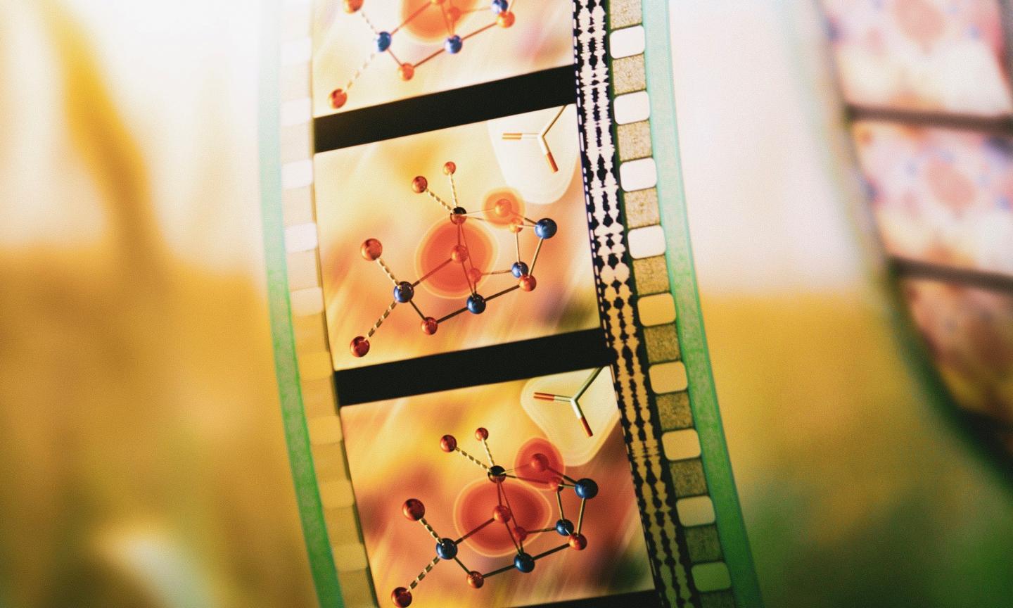
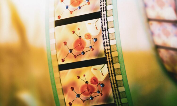
One of nature’s most important chemical reactions is now being captured in a breakthrough ‘molecular movie’

Credit: Greg Stewart/SLAC National Accelerator Laboratory
Using a unique combination of nanoscale imaging and chemical analysis, an international team of researchers has revealed a key step in the molecular mechanism behind the water splitting reaction of photosynthesis, a finding that could help inform the design of renewable energy technology.
“Life depends on the oxygen that plants and algae split from water; how they do it is still a mystery, but scientists, including our team, are slowly peeling away the layers to get to the answer,” said Vittal K. Yachandra, co-lead author of a new study published in PNAS and a chemist senior scientist at the Department of Energy’s (DOE) Lawrence Berkeley Laboratory (Berkeley Lab). “If we can understand this step of natural photosynthesis, it would enable us to use those design principles for building artificial photosynthetic systems that produce clean and renewable energy from sunlight and water.”
With an instrument that the team designed and fabricated, they analyzed photosynthetic proteins using both X-ray crystallography and X-ray emission spectroscopy. This dual approach, which the team pioneered and have been refining for the past 10 years, generates chemical and protein structure information from the same sample at the same time. The imaging was performed with the X-ray free-electron laser (XFEL) at the LCLS at SLAC National Laboratory, and at SACLA in Japan.
“With this technique, we get the overall picture of how the entire protein structure dynamically changes and we see the chemical intricacies occurring at the reaction site,” said co-lead author Junko Yano, a chemist senior scientist in Berkeley Lab’s Molecular Biophysics and Integrated Bioimaging (MBIB) Division. “The X-ray free electron laser produces extremely bright, short bursts of X-rays that allow us to not only analyze a protein at room temperature, which is how these reactions occur in nature, but also capture various moments over the reaction time scale.”
Traditional crystallography methods often require the sample proteins to be frozen; consequently, they can only generate snapshots of static proteins. This limitation makes it difficult for scientists to get a handle on how proteins actually behave in living organisms, because the molecules morph between different physical states during chemical reactions.
“The water-splitting reaction in photosynthesis is a cyclical process that needs four photons and cycles between four stable ‘states,'” said Yano. “Previously, we could only take pictures of these four states. But by taking multiple snapshots in time, we now can visualize how one state goes to the other.”
“We saw, really nicely, how the structure changes step-by-step as it transforms from one state to the next state,” said Jan F. Kern, MBIB chemist and co-author. “It is pretty exciting, because we can see the ’cause and effect’ and the role that each moving atom plays in this transition.”
Nicholas K. Sauter, co-author and MBIB computational senior scientist, added: “Essentially, we’re trying to take a ‘movie’ of a chemical reaction. We made a lot of progress to get to this point, in terms of our technology and our computational analyses. The work of our co-author Paul Adams and others in MBIB was critical to interpreting the XFEL and X-ray data. But we still have to get the other frames to see how the reaction is completed and the enzyme is ready for the next cycle.”
The Berkeley Lab researchers hope to continue the project once the many research sites that the entire international team relies upon – located in the U.S., Japan, Switzerland, and South Korea – are operating normally following the COVID-19 pandemic.
Kern concluded by noting that the technological milestone presented in this paper benefited greatly from the diverse expertise of the authors from SLAC, Uppsala and Umeå Universities in Sweden, Humboldt University in Germany, and from the capabilities of five DOE Office of Science user facilities: the Stanford Synchrotron Radiation Lightsource and LCLS at Stanford University, and the Advanced Light Source, Energy Sciences Network, and National Energy Research Scientific Computing Center at Berkeley Lab.
###
Other Berkeley Lab scientists who contributed to this work include: Ruchira Chatterjee, Louise Lassalle, Kyle D. Sutherlin, Iris D. Young, Sheraz Gul, In-Sik Kim, Philipp S. Simon, Isabel Bogacz, Cindy C. Pham, Nicholas Saichek, Trent Northen, Asmit Bhowmick, Robert Bolotovsky, Derek Mendez, Nigel W. Moriarty, James M. Holton, Aaron S. Brewster, and David Skinner.
This research was supported primarily by the DOE Office of Science and grants from the National Institutes of Health.
Founded in 1931 on the belief that the biggest scientific challenges are best addressed by teams, Lawrence Berkeley National Laboratory and its scientists have been recognized with 13 Nobel Prizes. Today, Berkeley Lab researchers develop sustainable energy and environmental solutions, create useful new materials, advance the frontiers of computing, and probe the mysteries of life, matter, and the universe. Scientists from around the world rely on the Lab’s facilities for their own discovery science. Berkeley Lab is a multiprogram national laboratory, managed by the University of California for the U.S. Department of Energy’s Office of Science.
DOE’s Office of Science is the single largest supporter of basic research in the physical sciences in the United States, and is working to address some of the most pressing challenges of our time. For more information, please visit energy.gov/science.
Media Contact
Aliyah Kovner
[email protected]
Related Journal Article
http://dx.


