• HOME
  • NEWS
  • EXPLORE
    • CAREER
      • Companies
      • Jobs
    • EVENTS
    • iGEM
      • News
      • Team
    • PHOTOS
    • VIDEO
    • WIKI
  • BLOG
  • COMMUNITY
    • FACEBOOK
    • INSTAGRAM
    • TWITTER
Saturday, February 7, 2026
BIOENGINEER.ORG
No Result
View All Result
  • Login
  • HOME
  • NEWS
  • EXPLORE
    • CAREER
      • Companies
      • Jobs
        • Lecturer
        • PhD Studentship
        • Postdoc
        • Research Assistant
    • EVENTS
    • iGEM
      • News
      • Team
    • PHOTOS
    • VIDEO
    • WIKI
  • BLOG
  • COMMUNITY
    • FACEBOOK
    • INSTAGRAM
    • TWITTER
No Result
View All Result
Bioengineer.org
No Result
View All Result
Home NEWS Science News Biology

Images of ‘invisible’ holes on cells may jumpstart research

Bioengineer by Bioengineer
March 3, 2020
in Biology
Reading Time: 3 mins read
0
IMAGE
Share on FacebookShare on TwitterShare on LinkedinShare on RedditShare on Telegram

IMAGE

Credit: Furukawa lab/CSHL, 2020


High blood pressure, inflammation, and the sensation of pain may rely in part on tiny holes on the surface of cells, called pores. Living cells react to the environment, often by allowing water and other molecules to pass through the cell’s surface membrane. Protein-based pores control this flow.

One set of pores, called pannexins, are like tiny “black holes” of biology; their existence has been confirmed by computational and functional evidence but until very recently, they were never actually seen. They help cells communicate with and react to their surroundings, but until scientists see what they look like, it is hard to know how. Now, a team of researchers from Cold Spring Harbor Laboratory (CSHL) and Cornell University have developed very high-resolution images of these elusive structures.

“We all knew these pores had to exist… but there’s definitely been some mystique about pannexins,” said postdoctoral researcher Kevin Michalski, who co-led the project. “Without images, it’s been really difficult to figure out exactly what these pores are doing.”

Michalski spent more than six years trying to get a clear image of pannexins.

“I had spent so long trying to set up experiments without really being able to see what I was working with. That’s what makes this so exciting,” Michalski explained. “For the first time, we can definitely see this detailed structure–it’s all new information.”

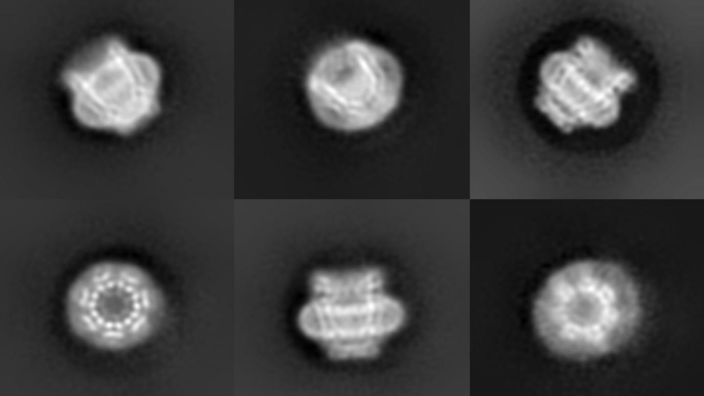
Michalski and Johanna Syrjanen, also in the Furukawa lab at CSHL collaborated with the Kawate lab at Cornell University, and published their findings in the journal eLife. They used a technique called cryo-electron microscopy (cryo-EM) to capture a half-million images of a pannexin pore, called “pannexin 1,” from many different angles. They then merged the images to form a three-dimensional model. Pannexin 1 is made up of seven identical proteins in a donut-like ring around a central pore.

Watch a video of the pannexin 1 model: https://www.youtube.com/watch?v=uvYjnHa_LPA

Researchers have hypothesized that pannexin 1 is responsible for releasing the signals that trigger a failing cell’s self-destruction. These signals prompt macrophages to destroy the ailing cell, clearing the way for new cells to thrive. Without the replacement of ailing cells with new cells, organisms age.

“But what’s interesting is that even cells that don’t go through the process of self-destruction can have pannexins,” Michalski said. “That means that there has to be some other important mechanisms that this pore is performing.”

“The way pannexin 1 is shaped on a cell is different from another kind of pore that my lab recently revealed,” added Professor Hiro Furukawa, who oversaw the research. “It appears that there are more kinds of pores than scientists originally anticipated. How and when they open and close are important questions to be explored.”

The flow of water, salts, and other molecules into a cell across a pore regulates the cell’s size. Cells that misregulate their size or water flow could contribute to high blood pressure, pain, and inflammation. Pannexins are also thought to allow much larger molecules to pass through their pores. Understanding how molecules negotiate their way through a pannexin could lead to new classes of drugs.

###

Media Contact
Sara Roncero-Menendez
[email protected]
516-367-6866

Original Source

https://www.cshl.edu/images-of-invisible-holes-on-cells-may-jumpstart-research/

Related Journal Article

http://dx.doi.org/10.7554/eLife.54670

Tags: BiologyCell Biology
Share12Tweet8Share2ShareShareShare2

Related Posts

Florida Cane Toad: Complex Spread and Selective Evolution

Florida Cane Toad: Complex Spread and Selective Evolution

February 7, 2026
New Study Uncovers Mechanism Behind Burn Pit Particulate Matter–Induced Lung Inflammation

New Study Uncovers Mechanism Behind Burn Pit Particulate Matter–Induced Lung Inflammation

February 6, 2026

DeepBlastoid: Advancing Automated and Efficient Evaluation of Human Blastoids with Deep Learning

February 6, 2026

Navigating the Gut: The Role of Formic Acid in the Microbiome

February 6, 2026
Please login to join discussion

POPULAR NEWS

  • Robotic Ureteral Reconstruction: A Novel Approach

    Robotic Ureteral Reconstruction: A Novel Approach

    82 shares
    Share 33 Tweet 21
  • Digital Privacy: Health Data Control in Incarceration

    63 shares
    Share 25 Tweet 16
  • Study Reveals Lipid Accumulation in ME/CFS Cells

    57 shares
    Share 23 Tweet 14
  • Breakthrough in RNA Research Accelerates Medical Innovations Timeline

    53 shares
    Share 21 Tweet 13

About

We bring you the latest biotechnology news from best research centers and universities around the world. Check our website.

Follow us

Recent News

Evaluating Pediatric Emergency Care Quality in Ethiopia

TPMT Expression Predictions Linked to Azathioprine Side Effects

Improving Dementia Care with Enhanced Activity Kits

Subscribe to Blog via Email

Success! An email was just sent to confirm your subscription. Please find the email now and click 'Confirm' to start subscribing.

Join 73 other subscribers
  • Contact Us

Bioengineer.org © Copyright 2023 All Rights Reserved.

Welcome Back!

Login to your account below

Forgotten Password?

Retrieve your password

Please enter your username or email address to reset your password.

Log In
No Result
View All Result
  • Homepages
    • Home Page 1
    • Home Page 2
  • News
  • National
  • Business
  • Health
  • Lifestyle
  • Science

Bioengineer.org © Copyright 2023 All Rights Reserved.