Credit: Courtesy of Van Andel Institute
GRAND RAPIDS, Mich. (Nov. 6, 2019) — Scientists have devised a technique to sort out which heart cells can replicate and which cannot, a critical step toward treatments that may one day help the heart heal itself after injury.
The method, published in the journal Circulation Research, removes a significant roadblock to developing ways to regrow healthy cardiac muscle tissue, a feat not currently possible.
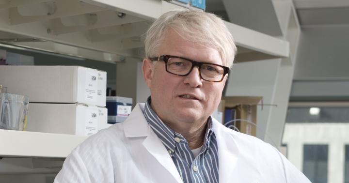
“This new technique solves a longstanding problem that for years has stymied our ability to develop regenerative treatments for the heart,” said Stefan Jovinge, M.D., Ph.D., the study’s senior author and director of the DeVos Cardiovascular Research Program at Van Andel Institute and Spectrum Health. “It’s a major step forward that we aim to translate into improved patient care.”
For decades, scientists have searched for ways to harness the heart’s regenerative potential to fix damage related to heart attack and heart failure, but their attempts have been largely unsuccessful.
Unlike the skin or bones, which readily heal by stitching together wounds or breaks with new, healthy cells, heart muscle cells largely lose their ability to replicate early in life (instead, they progress through the cell cycle but, in most cases, they do not actually divide). This leaves patients and physicians with only a few surgical options to mitigate further damage and just one option to totally repair the problem should the damage be too severe: a heart transplant.
The new method combines two technologies — molecular beacon technology and fluorescence activated cell-sorting — to specifically isolate cells that successfully divide. This will allow scientists to determine the mechanisms underlying heart muscle cells’ regenerative potential, which is critical to the development of regenerative strategies aimed to cure patients with heart injury.
“Now that we can accurately identify these cells, we can start to determine the mechanisms that allow them to divide and develop ways to jump-start this process,” Jovinge said. “This work is an excellent example of how basic research can have a major impact on future clinical care.”
The findings build on previous research by Jovinge’s team that showed, contrary to long-held beliefs, some cells in the heart are indeed capable of replicating. This discovery was hailed as a “notable advance of 2015” by Nature Medicine, one of the world’s top medical research journals.
In addition to Jovinge, authors include Hsiao-yun Y. Milliron, Ph.D., Matthew J. Weiland, M.S., and Eric J. Kort, M.D., M.S. of the DeVos Cardiovascular Research Program, a joint effort between Van Andel Institute and Spectrum Health. Van Andel Institute’s Bioinformatics and Biostatistics Core, Genomics Core and Optical Imaging Core also contributed to this work.
Research reported in this publication was supported by the Richard and Helen DeVos Foundation. The content is solely the responsibility of the authors and does not necessarily represent the official views of any funding organization.
###
ABOUT VAN ANDEL INSTITUTE
Van Andel Institute (VAI) is committed to improving the health and enhancing the lives of current and future generations through cutting edge biomedical research and innovative educational offerings. Established in Grand Rapids, Michigan, in 1996 by the Van Andel family, VAI is now home to more than 400 scientists, educators and support staff, who work with a growing number of national and international collaborators to foster discovery. The Institute’s scientists study the origins of cancer, Parkinson’s and other diseases and translate their findings into breakthrough prevention and treatment strategies. Our educators develop inquiry-based approaches for K-12 education to help students and teachers prepare the next generation of problem-solvers, while our Graduate School offers a rigorous, research-intensive Ph.D. program in molecular and cellular biology.
Media Contact
Beth Hinshaw Hall
[email protected]
616-234-5519
Related Journal Article
http://dx.