In a groundbreaking advancement in head and neck surgery, researchers have harnessed cutting-edge three-dimensional (3D) reconstruction technology combined with transoral endoscopic techniques to significantly enhance the precision in the removal of parapharyngeal space tumors. This novel approach represents a major leap forward in the surgical treatment of these complex tumors, which are notoriously difficult to access due to their proximity to critical anatomical structures.
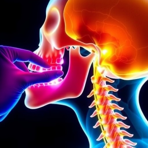
The parapharyngeal space, a deep compartment adjacent to vital neurovascular bundles and the upper aerodigestive tract, poses substantial challenges for surgeons. Traditional surgical approaches often involve extensive incisions and carry a heightened risk of complications, including injury to nerves and blood vessels. By integrating advanced 3D reconstruction based on detailed medical imaging, surgeons can now create intricate digital models of both the tumor and its surrounding anatomical environment, offering an unparalleled roadmap prior to the operation.
This technology was applied in a recent report involving three patients diagnosed with different types of parapharyngeal tumors—a hemangioma, a nerve sheath tumor, and a pleomorphic adenoma. By transforming the patients’ imaging data into precise, manipulable 3D models, the surgical teams could visualize tumor boundaries, their relation to neighboring tissues, and tailor the operative strategy accordingly. This preoperative planning optimized the choice of incision sites and minimized disruption to the surrounding musculature and neurovascular structures.
Utilizing general anesthesia with transoral endoscopic assistance, these surgeries involved accessing tumors through the lateral palatoglossal arch and pharyngo-palatine muscular space, anatomical corridors rarely exploited in traditional methods. The endoscope’s enhanced visualization allowed surgeons to operate with heightened accuracy in a minimally invasive manner, reducing trauma to the patient and expediting postoperative recovery.
Post-surgical evaluations confirmed the initial diagnoses: hemangioma in the left parapharyngeal space, a complex nerve sheath tumor on the right, and pleomorphic adenoma on the left. All three patients demonstrated significant relief of symptoms after their respective surgeries, with no postoperative complications reported. These outcomes strongly underscore the safety, efficacy, and patient benefits conferred by combining 3D visualization and endoscopic guidance.
Beyond clinical outcomes, this integration of 3D imaging and transoral endoscopy heralds a new paradigm in personalized surgical care. By providing surgeons with immersive, interactive models, this approach reduces intraoperative uncertainties, limits healthy tissue excision, and preserves critical structures. Such precision is especially crucial in the parapharyngeal space, where millimeters can differentiate between successful resection and debilitating complications.
Moreover, the minimally invasive nature of this technique corresponds with shorter hospital stays and quicker return to normal function, contributing to overall improvements in healthcare quality and cost efficiency. Patients experience less postoperative pain, lower infection risks, and reduced scarring compared to conventional open procedures, making this a patient-centric advancement in otolaryngology and surgical oncology.
The incorporation of cutting-edge software for three-dimensional reconstruction also opens new avenues in medical education and surgical training. Surgeons-in-training can engage with virtual models to understand tumor anatomy and rehearse operative maneuvers in a simulated environment, elevating the standard of surgical preparedness without patient risk.
While this study focused on benign tumors of the parapharyngeal space, the implications extend to a broad spectrum of complex head and neck lesions. As imaging modalities and computational algorithms continue to evolve, future surgical interventions could be increasingly personalized, targeted, and minimally invasive.
In conclusion, the synergistic use of medical imaging-derived 3D reconstruction and transoral endoscopic surgery represents a transformative step forward in the management of parapharyngeal space tumors. By enabling surgeons to visualize, strategize, and execute tumor resections with unmatched precision and safety, this approach promises to redefine standards of care, reduce patient morbidity, and inspire further innovations in surgical technology.
This advancement exemplifies the power of integrating digital healthcare tools with traditional surgical expertise, paving the way for a future where complex tumor resections become safer, faster, and more effective. As this technique gains wider adoption, it could significantly improve quality of life for countless patients facing challenging head and neck tumors.
As research progresses, multidisciplinary collaboration between biomedical engineers, radiologists, and surgeons will be essential to refine these technologies and expand their applicability. The future of surgical oncology lies at the intersection of innovation and precision, with 3D technology and endoscopy leading the charge.
Subject of Research: Surgical treatment of parapharyngeal space tumors using three-dimensional reconstruction and transoral endoscopic assistance.
Article Title: 3D technology facilitates transoral endoscopic precision surgery of parapharyngeal space tumors.
Article References:
Shi, Y., Han, S., Guo, R. et al. 3D technology facilitates transoral endoscopic precision surgery of parapharyngeal space tumors. BioMed Eng OnLine 24, 136 (2025). https://doi.org/10.1186/s12938-025-01469-3
Image Credits: AI Generated
DOI: 17 November 2025
Tags: 3D reconstruction technology in surgeryadvanced medical imaging for surgical planninganatomical modeling in head and neck surgerychallenges in parapharyngeal space surgerieshemangioma nerve sheath tumor treatmentinnovative approaches in oncology surgeryintegration of technology in surgical practiceminimizing complications in tumor surgeryparapharyngeal tumor removal techniquespleomorphic adenoma surgical strategiesprecision surgery for complex tumorstransoral endoscopic surgery advancements

