In an era where advanced medical technology synergizes with innovative engineering solutions, the advent of three-dimensional (3D) printing has opened new frontiers in the management of complex congenital heart defects. Among these conditions, double outlet right ventricle (DORV) poses unique challenges to clinicians striving to optimize patient outcomes. Traditionally, treatment strategies for DORV have been fraught with difficulty, often requiring invasive procedures and lengthy surgeries. However, recent advancements in three-dimensional modeling and printing have heralded a new age of patient-specific surgical planning, facilitating a more tailored approach to this intricate condition.
DORV is characterized by a congenital anatomical anomaly where both great arteries, the aorta and pulmonary artery, arise from the right ventricle. This condition can lead to severe hemodynamic derangements if left uncorrected. The presence of an associated ventricular septal defect (VSD) complicates matters further, particularly when the VSD is positioned remotely from the arterial roots. Surgical intervention aims to reconfigure the heart’s architecture, enabling proper separation of oxygenated and deoxygenated blood. With the aid of 3D printing, preliminary modeling provides invaluable insights into these complex anatomical configurations, granting surgeons enhanced visualization prior to any cuts being made.
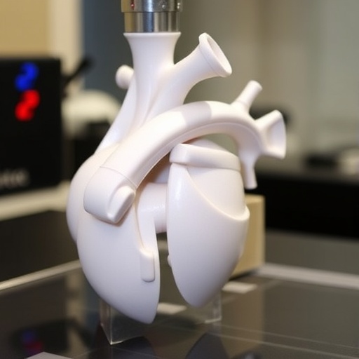
3D printing functions as a transformative tool within the surgical landscape by creating anatomically accurate models of a patient’s heart structure derived from imaging studies such as MRI or CT scans. These models allow for detailed preoperative planning, enabling cardiac surgeons to practice the procedure on an exact replica of the patient’s anatomy. This pre-surgical rehearsal fosters familiarity with the unique challenges posed by DORV, improving the efficiency and safety of the actual surgery. As a result, the risk of complications can be substantially reduced, leading to better postoperative recovery trajectories.
Moreover, the incorporation of 3D-printed models into clinical practice facilitates comprehensive discussions among multidisciplinary teams, including cardiologists, surgeons, and radiologists. The shared use of visual aids can enhance collaborative decision-making, ensuring that every aspect of patient care is meticulously thought out. The clarity provided by tangible models helps bridge the gaps between complex anatomical concepts and patient understanding, paving the way for more informed consent processes. Patients and families often find comfort in visualizing procedures, ultimately leading to improved satisfaction and adherence to proposed treatment plans.
Beyond the immediate surgical applications, the implications of 3D printing extend into the realm of training and education for new and seasoned cardiothoracic surgeons alike. Surgical education often relies on cadaveric studies, which, while valuable, lack the specificity and real-time interaction that 3D models can provide. With the aid of 3D printing, training scenarios can be tailored to address particular pathologies, ensuring that practitioners are well-prepared for the diverse challenges they may encounter in live surgical settings.
As we explore the advancements, it’s crucial to acknowledge the ethical considerations and financial implications associated with implementing 3D printing in cardiac surgeries. The cost of high-quality 3D printing technology can be a barrier in some healthcare settings. However, as the technology becomes more ubiquitous and accessible, economies of scale may lead to a decrease in costs, allowing for broader adoption in clinics across various demographics.
Further research is warranted to quantify the long-term impacts of 3D printing on surgical outcomes in patients with DORV. Clinical trials should be designed to evaluate both morbidity and mortality rates, as well as patient quality of life metrics following interventions facilitated by preoperative modeling. The iterative learning process inherent in research will allow clinicians to refine techniques and push the boundaries of what is clinically feasible.
Moreover, the possibilities of future directions in 3D printing raise exciting prospects. Innovations such as bioprinting, where viable tissues or organs are printed, offer the tantalizing potential to address current limitations in donor organ availability. Although still in the nascent phases of research, the vision of printing heart structures tailored to individual patients may someday transform the landscape of transplantation and regenerative medicine.
In conclusion, the use of 3D printing technology in the management of double outlet right ventricle represents a significant stride towards personalized medicine. By employing tailored approaches through accurate and patient-specific models, clinicians can optimize surgical outcomes and improve patient experiences. As research progresses and technological advancements continue to unfold, it is not just the individual patients with DORV who stand to benefit but the entire landscape of cardiac surgery itself.
As healthcare professionals remain committed to integrating cutting-edge technologies into patient care, the hopeful narrative of 3D printing illustrates the synergy of art and science—an evolving paradigm where intricate biological puzzles are met with innovative engineering solutions, ultimately pushing the frontiers of medical possibility.
Subject of Research: The role of three-dimensional printing in enhancing management of double outlet right ventricle.
Article Title: Enhancing management of double outlet right ventricle when the interventricular communication is remote from the arterial roots through three-dimensional printing.
Article References:
Kindi, H.N.A., Maddali, M.M., Kandachar, P.S. et al. Enhancing management of double outlet right ventricle when the interventricular communication is remote from the arterial roots through three-dimensional printing. 3D Print Med 11, 18 (2025). https://doi.org/10.1186/s41205-025-00265-y
Image Credits: AI Generated
DOI: https://doi.org/10.1186/s41205-025-00265-y
Keywords: double outlet right ventricle, congenital heart defects, 3D printing, surgical planning, patient-specific models, cardiac surgery, hemodynamics, bioprinting.


