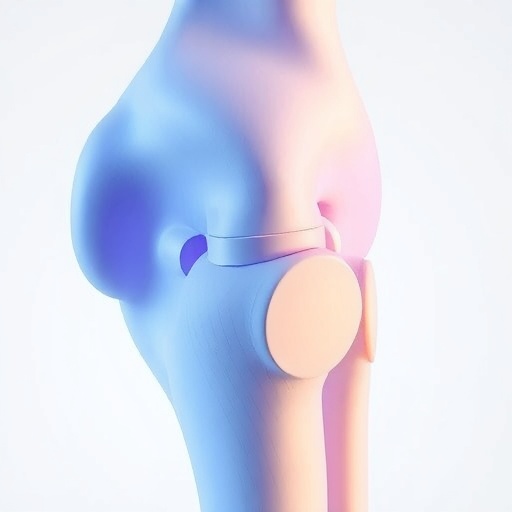
In a groundbreaking study conducted by Yan, Yao, and Liu, researchers delve deep into the intricate relationship between the three-dimensional (3D) shape of the patella, commonly known as the kneecap, and the propensity for patellar dislocation. This fascinating topic is garnering increasing attention in the fields of biomechanics and orthopedics, particularly due to the rising incidents of knee injuries among athletes and active populations. The significance of understanding the anatomical variations that may predispose individuals to such injuries cannot be overstated, as it has the potential to inform prevention strategies and treatment protocols.
The study harnesses advanced imaging techniques and statistical shape modeling to investigate the geometric features of the patella. Utilizing automated coordinate algorithms, the researchers gather and analyze data from a variety of subjects, enabling them to model the patellar shape with unprecedented precision. This methodological approach marks a substantial leap forward in the ability to quantify and interpret the complexities of patellar morphology. By doing so, the authors aim to provide clearer insights into how specific 3D shapes may influence the likelihood of dislocation events.
Patellar dislocation occurs when the patella shifts out of its normal alignment within the femoral groove, and its incidence is often associated with sports activities that involve sudden changes in direction or intense physical impact. Individuals who have experienced a patellar dislocation frequently exhibit recurrent instability in the knee joint, leading to a cycle of injuries that can significantly affect both athletic performance and overall quality of life. Therefore, identifying predisposing anatomical features through this study may vastly improve rehabilitation protocols and preventive measures for these individuals.
In their analysis, the authors collect a substantial dataset that includes a diverse range of patellar shapes from different age groups and activity levels. This diversity is crucial, as it enables the researchers to build a comprehensive model that accurately represents the variations encountered in clinical practice. The automated algorithms employed facilitate the smooth processing of this data, ensuring that the study’s findings are both robust and reproducible. As a result, the study provides a valuable framework for understanding how geometric variables correlate to dislocation risk.
Crucially, the study reveals that certain shapes of the patella are more susceptible to dislocation than others, leading to nascent discussions about personalized medicine in orthopedic treatment. Clinicians, equipped with new insights from statistical shape modeling, may be better positioned to assess individual risk factors and tailor rehabilitation strategies accordingly. This shift towards personalized approaches offers the promise of delivering more effective treatments and reducing the incidence of patellar dislocation among vulnerable populations.
Alongside its clinical implications, this research also underlines the importance of interdisciplinary collaboration in modern scientific inquiries. By combining expertise from advanced imaging, computational modeling, and clinical orthopedics, Yan, Yao, and Liu exemplify how collaborative efforts can drive significant advancements in understanding complex medical issues. The application of technology in traditional medicine fields not only enhances the quality of research but also paves the way for innovative solutions that can address longstanding healthcare challenges.
Furthermore, the researchers impressively manage to balance technical rigor with accessibility in the publication of their findings. By clearly articulating their methods and results, they ensure that their work is not only suitable for specialists but also relevant to a broader audience. This accessibility is essential in fostering a well-informed public discourse around knee health and injuries, making it an exemplar of how scientific research should be communicated.
Looking forward, it becomes increasingly evident that the implications of this study extend beyond the immediate scope of patellar dislocation. The analytical framework developed by the researchers could be applied to other musculoskeletal disorders, potentially illuminating additional anatomical risk factors in similar injury-prone populations. Such a shift in perspective could catalyze further research, drawing attention to the importance of biological shape in sports medicine and rehabilitation.
In conclusion, the work by Yan, Yao, and Liu on the association between 3D patellar shape and patellar dislocation is a landmark contribution to the field of biomedical engineering and orthopedics. By employing sophisticated modeling techniques and comprehensive data analysis, the researchers have shed light on a previously obscure area of study, generating vital information that can help shape future approaches to injury prevention and management. As the field continues to evolve, the integration of new technologies with clinical practices stands to revolutionize orthopedic care and improve outcomes for countless individuals.
This study not only enhances our understanding of knee mechanics but also calls for ongoing research and discourse in the realm of biomechanics. As new technologies emerge, the potential to refine our understanding of human anatomy and its clinical implications grows exponentially, heralding a future where injuries can be treated more effectively and human movement is better understood.
As we consider the long-term ramifications of this research, it emphasizes the pressing need for continuous exploration of anatomical variations within the field. Understanding how different shapes and sizes influence biomechanical behaviors will ultimately lead to enhanced athletic performance and, more importantly, greater overall health and well-being for individuals across all activity levels.
Strong implications for clinical practice stem from this research, highlighting the importance of individualized assessment and treatment strategies in orthopedic care. By recognizing and addressing the unique anatomical characteristics of each patient, healthcare professionals can create more tailored rehabilitation programs that cater to specific needs, thus improving recovery outcomes and reducing the risk of future injuries.
Ultimately, as we advance our knowledge of the complex interplay between anatomy and biomechanics, studies like these serve as vital stepping stones in driving the orthopedic field forward, with the potential to reform approaches to injury prevention, management, and rehabilitation in the years to come.
Subject of Research: The association between 3D patellar shape and patellar dislocation
Article Title: 3D Patellar Shape is Associated with Patellar Dislocation: an Automated Coordinate Algorithm and Statistical Shape Modeling Analysis
Article References:
Yan, Y., Yao, J., Liu, Z. et al. 3D Patellar Shape is Associated with Patellar Dislocation: an Automated Coordinate Algorithm and Statistical Shape Modeling Analysis.
Ann Biomed Eng (2026). https://doi.org/10.1007/s10439-025-03970-1
Image Credits: AI Generated
DOI: https://doi.org/10.1007/s10439-025-03970-1
Keywords: Patellar dislocation, 3D shape modeling, biomechanics, orthopedic research, injury prevention, personalized medicine, knee health.
Tags: 3D patellar shape analysisadvanced imaging techniques in orthopedicsanatomical variations in patellaautomated coordinate algorithms in researchbiomechanics of knee injuriesgeometric features of the patellaknee injury incidence among athletespatellar dislocation risk factorsprevention strategies for knee injuriesstatistical shape modeling in biomechanicstreatment protocols for dislocated patellaunderstanding patellar morphology

