A new imaging technique for real 3D vascular ultrasound could become a key tool in strategies aimed at preventing cardiovascular disease in apparently healthy persons, complementing traditional risk parameters such as cholesterol and high blood pressure. The new results, published in JACC: Cardiovascular Imaging, show that real 3D vascular ultrasound is reliable, accurate, and faster than previous methods for the assessment of plaque volume in the carotid and femoral arteries.
Credit: CNIC
A new imaging technique for real 3D vascular ultrasound could become a key tool in strategies aimed at preventing cardiovascular disease in apparently healthy persons, complementing traditional risk parameters such as cholesterol and high blood pressure. The new results, published in JACC: Cardiovascular Imaging, show that real 3D vascular ultrasound is reliable, accurate, and faster than previous methods for the assessment of plaque volume in the carotid and femoral arteries.
The burden, or quantity, of atherosclerosis in the carotid and femoral arteries is a well-established marker of cardiovascular risk and is highlighted as a key parameter in international clinical practice guidelines and expert consensus documents. There is therefore a recognized need for better and easy-to-use methods for measuring plaque burden that can be used as population screening tools.
The new imaging method was first validated and implemented in a study of almost 200 healthy participants with an intermediate cardiovascular risk from the Athero Brain: Head-to-Heart study, led by Dr. Valentín Fuster, Director General of the Centro Nacional de Investigaciones Cardiovasculares (CNIC). The method has now been incorporated into the PESA-CNIC-SANTANDER study, also led by Dr. Fuster, where it is being used to assess more than 4000 healthy individuals over a 9-year follow-up.
The PESA-CNIC-SANTANDER study, which started in 2010 and was recently extended until 2030, is one of the most important cardiovascular prevention studies in the world.
The CNIC researchers partnered with Philips Ultrasound and Philips Research Paris-Medisys to develop a new probe and software for real 3D ultrasound to facilitate exploration of the carotid and femoral arteries and speed up quantification of atherosclerotic plaque volume. As Dr. Fuster explained, “it is clear that traditional clinical evaluations based on measurements of cholesterol, blood pressure, blood glucose, and lifestyle habits cannot, on their own, accurately determine accumulated damage in the cardiovascular system, and without this crucial information we cannot take appropriate decisions to prevent acute events such as myocardial infarction or stroke.”
The key to personalized prevention and treatment strategies, added Dr. Fuster, “is the ability to detect and quantify an individual’s accumulated cardiovascular damage, or atherosclerotic burden, using noninvasive imaging techniques.”
The newly validated 3D vascular probe incorporates 3D matrix technology, which underpins the most advanced 3D ultrasound techniques. CNIC Clinical Research Director Dr. Borja Ibáñez explained that the new technology allows simultaneous analysis by 2D and 3D ultrasound, includes all functionalities (color doppler, power-doppler, and contrast ultrasound), and is easily incorporated into daily clinical practice by technical and medical teams already experienced in ultrasound, emphasizing that “the integrated analysis software incorporates real 3D data processing.”
In addition to demonstrating the accuracy of 3D matrix ultrasound, the study demonstrates that the new technique takes just half the time needed by previous methods to obtain all the information required for the definition of carotid and femoral plaque burden, essential information for correct patient management.
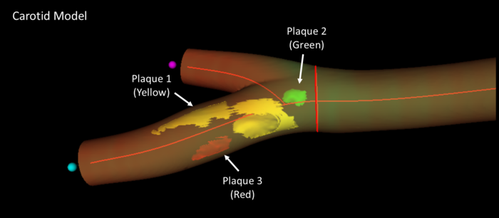
For patients, the outstanding feature of the new method is that the software generates a virtual 3D image of their own arteries, allowing them to see the accumulated damage. “When patients see the state of their arteries, this impresses upon them the need to change their lifestyle, in graphic manner not achieved by reading a list of analytical data,” said first author Dr. Beatriz López Melgar, a cardiologist at Hospital Universitario La Princesa and head of the 3D Cardioprevention Program at Hospital HM Montepríncipe in Madrid.
And for medical professionals, continued Dr. López Melgar, “the ability to view plaques in 3D allows us to assess them more precisely and in their enterity, something not possible with conventional 2D methods. 3D ultrasound also provides invaluable information about plaque morphology”.
“Likewise, this technological advance will soon allow us to analyze plaque composition and to use this information to assess the burden of ‘adverse plaques’ (plaques with a high lipid content that may be at increased risk of rupture and triggering events, such as stroke). Adverse plaque burden is a very promising marker that until now could only be assessed using highly advanced techniques that involve radiation, such as CAT and PET. Now, with 3D matrix technology, measuring adverse plaque burden is a realistic goal of cardiovascular ultrasound studies.”
For this reason, added Dr. López Melgar, “we believe that the development of ultrasound methods will contribute to the expansion of personalized medicine and the use of diagnostic imaging techniques, and will also help to ensure that improvements in patient care produce benefits for a larger sector of the population.”
López Melgar concluded that, with the development of this technology, “we now have a tool that can be used on-the-fly in an initial consultation, speeding up decision making—an important consideration in cardiovascular prevention, where time is of the essence.”
Collaborators on this project included scientists from the Spanish cardiovascular research network (CIBERCV), and financial support was provided by the Ministerio de Economía, Industria y Competividad (MEIC) and the European Regional Development Fund.
About the CNIC
The Centro Nacional de Investigaciones Cardiovasculares (CNIC), directed by Dr. Valentín Fuster, is dedicated to cardiovascular research and the translation of knowledge gained into real benefits for patients. The CNIC, recognized by the Spanish government as a Severo Ochoa center of excellence, is financed through a pioneering public-private partnership between the government (through the Carlos III Institute of Health) and the Pro-CNIC Foundation, which brings together 12 of the most important Spanish private companies.
Journal
JACC Cardiovascular Imaging
Method of Research
Randomized controlled/clinical trial
Subject of Research
Not applicable
Article Title
“New 3-Dimensional Volumetric Ultrasound Method for Accurate Quantification of Atherosclerotic Plaque Volume”
Article Publication Date
16-Mar-2022


