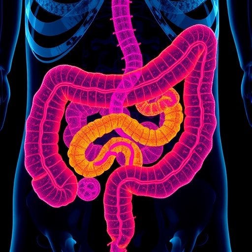
In a groundbreaking advancement poised to redefine gastrointestinal diagnostics, researchers have unveiled a novel three-dimensional imaging technique that offers unprecedented visualization of both healthy and inflamed intestinal tissues. This pioneering approach harnesses the power of optical coherence tomography (OCT), a cutting-edge optical imaging modality, to generate detailed, ex-vivo images of the small intestine and colon, bringing new clarity to the complex architecture and pathology of these vital organs.
Optical coherence tomography operates on principles akin to ultrasound imaging but utilizes light waves instead of sound. By measuring the echo time delay and intensity of backscattered light from tissue microstructures, OCT constructs high-resolution cross-sectional images. The research team employed a sophisticated OCT system optimized for tissue penetration and resolution, enabling them to visualize the intricate layers of intestinal walls with microscopic precision. This level of structural detail is critical, as subtle morphological changes in gut tissue often signify the onset or progression of inflammatory diseases.
Historically, the evaluation of intestinal inflammation has relied heavily on invasive endoscopic biopsies and histopathological examination, which, while informative, offer limited spatial context and can be hampered by sampling bias. The emergence of OCT as a non-destructive imaging technique represents a paradigm shift, providing volumetric data that captures the three-dimensional complexity of tissue architecture without the need for extensive tissue processing. This not only accelerates diagnostic workflows but also preserves tissue integrity for subsequent analyses.
In their study, the researchers meticulously prepared ex-vivo segments of normal and inflamed small intestine and colon tissue, replicating conditions pertinent to common inflammatory disorders such as Crohn’s disease and ulcerative colitis. Imaging these specimens with OCT revealed distinct morphological differences, including variations in mucosal thickness, crypt architecture, and submucosal changes, which are hallmark indicators of inflammation severity. These morphological signatures were rendered in vivid three-dimensional reconstructions, offering a holistic view that surpasses traditional two-dimensional histology.
The technical prowess of the OCT system was evident in its axial and lateral resolution capabilities, which achieved micrometer-scale detail, essential for discerning the subtle pathological alterations within the mucosal and submucosal layers. This precision allowed the team to identify features such as mucosal erosion, crypt abscesses, and localized edema, all of which are crucial for accurate disease staging and therapeutic decision-making. Moreover, the label-free nature of OCT obviates the need for exogenous contrast agents, streamlining the imaging process and reducing potential artifacts.
Beyond static imaging, the volumetric data acquired through OCT facilitate quantitative analyses of tissue morphology. Parameters such as crypt density, wall thickness, and inflammation indices can be algorithmically extracted, paving the way for the development of automated diagnostic tools. This integration of imaging and computational analytics represents an exciting frontier in digital pathology, potentially enabling real-time, objective assessment of gastrointestinal health in clinical settings.
The implications of this research extend beyond diagnostics. The capability to non-invasively monitor the dynamic progression of intestinal inflammation ex-vivo offers valuable insights for drug development and therapeutic evaluation. Researchers can now visualize how candidate treatments modulate tissue structure at a microscale level, accelerating the translation of novel anti-inflammatory agents from bench to bedside. This technology also opens avenues for personalized medicine approaches, tailoring therapies based on detailed morphological phenotyping of patient tissue samples.
Importantly, the use of ex-vivo human tissues in these experiments underscores the translational potential of OCT imaging. While in vivo applications face challenges such as motion artifacts and limited optical penetration due to living tissue scattering, technological advancements including catheter-based OCT probes and adaptive optics are rapidly addressing these hurdles. The present work lays a critical foundation for future in vivo imaging endeavors, promising less invasive, higher-resolution gastrointestinal diagnostics.
This study also contributes to a deeper understanding of gut tissue pathology by providing a direct comparison between normal and inflamed states within the same imaging platform. Such consistency ensures that observed differences are attributable to pathological processes rather than inter-modality variability. The comprehensive three-dimensional datasets generated could inform new criteria for disease classification and severity grading, refining clinical guidelines and improving patient outcomes.
From a broader technological perspective, this investigation illustrates the versatility of OCT beyond its traditional ophthalmologic applications. The expansion of OCT into gastroenterology exemplifies interdisciplinary innovation, where advancements in photonics and computational imaging synergize with clinical needs. As OCT instrumentation continues to evolve, incorporating enhancements such as higher-speed scanning and multimodal imaging integration, its utility in comprehensive tissue characterization is poised to expand further.
The researchers also addressed challenges inherent in large volumetric data handling and visualization. By implementing advanced image processing algorithms and three-dimensional rendering techniques, they ensured that the complex information derived from OCT scans could be intuitively interpreted by clinicians and researchers alike. This emphasis on user-friendly visualization tools is critical for widespread adoption of OCT imaging technologies in clinical workflows.
Looking ahead, the study’s authors envision integrating OCT with other imaging and molecular analysis techniques to create multimodal platforms capable of simultaneously providing structural, functional, and biochemical information. Such holistic approaches could revolutionize gastrointestinal medicine by enabling a multi-faceted assessment of intestinal health, advancing the precision and scope of diagnostics.
Moreover, the adoption of this technology in routine clinical practice could significantly reduce healthcare costs and patient burden by minimizing invasive procedures. Early detection of intestinal inflammation translates to timely interventions, potentially mitigating disease progression and associated complications. OCT’s rapid acquisition times and real-time imaging potential further enhance its appeal as a diagnostic adjunct.
In summary, the innovative use of optical coherence tomography for three-dimensional ex-vivo visualization of small intestinal and colonic tissues represents a major leap forward in gastrointestinal pathology. By delivering high-resolution, volumetric images that differentiate normal from inflamed tissue states, this technique offers a powerful, non-destructive alternative to conventional histopathology. As OCT technology matures and transitions toward in vivo applications, it holds promise to transform the diagnosis, monitoring, and treatment of inflammatory bowel diseases and beyond.
This research epitomizes the synergy between photonics, biomedical engineering, and clinical medicine, heralding a new era where detailed tissue imaging informs patient care with unprecedented clarity and precision. Ultimately, such innovations underscore the potential of optical imaging technologies to unravel complex biological systems and improve health outcomes on a global scale.
Subject of Research: Three-dimensional ex-vivo visualization of normal and inflamed small intestine and colonic tissue using optical coherence tomography.
Article Title: Three-dimensional ex-vivo visualization of normal and inflamed small intestine and colonic tissue using optical coherence tomography.
Article References:
Matt, A., Li, Y., Song, A. et al. Three-dimensional ex-vivo visualization of normal and inflamed small intestine and colonic tissue using optical coherence tomography. Sci Rep (2026). https://doi.org/10.1038/s41598-026-46293-4
Image Credits: AI Generated
Tags: 3D ex-vivo intestinal imagingadvanced optical imaging modalities for tissuedetailed visualization of small intestine and colonhigh-resolution OCT in gastrointestinal diagnosticsmicroscopic precision in gut tissue imagingnon-invasive imaging of intestinal inflammationnovel techniques in gastrointestinal pathologyOCT imaging for inflammatory bowel diseaseOCT versus endoscopic biopsyoptical coherence tomography for gut analysisoptical imaging of healthy and diseased intestinesvolumetric imaging of intestinal walls


