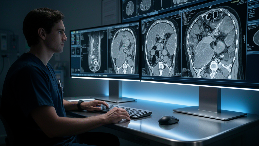
Radboud University Medical Centre researchers have developed a modified 3D fullres nnU-net neural network capable of detecting and segmenting traumatic liver injuries on abdominal computed tomography scans with high diagnostic accuracy. In an evaluation across twenty test cases, the multi-task architecture achieved an area under the receiver operating characteristic curve of 0.91, alongside an overall classification accuracy, sensitivity, and specificity of 0.90, while attaining a voxel-level Dice similarity coefficient of 0.779 for traumatic parenchymal lesion segmentation. The retrospective investigation, structured under STROBE reporting guidelines and conducted under formal institutional review board approval, represents a foundational proof-of-principle engineering initiative designed to support rapid patient triage in high-stress emergency departments and military medical units. By executing both injury classification and volumetric segmentation within a single computational pipeline, the artificial intelligence architecture directly addresses diagnostic bottlenecks that arise during initial trauma surveys, where clinicians must interpret complex multi-detector scans under physical fatigue and pressing time constraints.
Abdominal trauma accounts for approximately ten percent of all physical injuries sustained during combat operations, whereas within civilian emergency care, the liver represents the most frequently injured solid abdominal organ. Owing to its dense vascular network and massive physiological blood flow, mechanical disruption of the hepatic parenchyma poses an immediate risk of major hemorrhage, systemic morbidity, and rapid mortality. While computed tomography serves as the gold standard for assessing suspected hepatic trauma due to its superior spatial resolution and diagnostic sensitivity, interpreting these complex imaging datasets in emergent clinical environments introduces substantial logistical hurdles. Triage clinicians must balance clinical risks under severe resource scarcity, particularly when managing multi-casualty events or combat casualties in austere conditions. Research cited by the authors demonstrates that diagnostic interpretive errors occur more frequently within emergency departments than across other hospital divisions, driven by incomplete patient information, fatigue, and stressful multidisciplinary workflows. In military operational settings, these diagnostic challenges are further compounded by ongoing hostile threats and scarce imaging infrastructure.
To develop and validate the deep learning model, the research team retrospectively reviewed trauma records from Radboud University Medical Centre, a Level One trauma centre and military-affiliated hospital in Nijmegen, Netherlands. Eligible participants were patients aged sixteen and older who sustained high energetic trauma between 2015 and 2021 and underwent abdominal computed tomography less than twenty-four hours post-trauma. Under institutional policy, all patients consented to data use. The analytical dataset comprised 108 patients, divided into 54 individuals with documented traumatic liver injury and 54 uninjured controls. The injured cohort had a mean age of 37.1 years with 63 percent males, whereas uninjured controls averaged 52.6 years with 70.4 percent males. Traffic accidents formed the primary trauma mechanism in both groups, accounting for 70.4 percent of injured cases and 63 percent of controls, followed by falls and sharp object injuries. Mean injury severity score reached 24.8 in injured patients compared to 18.3 among uninjured controls.
All diagnostic scans were acquired on an Aquilion ONE computed tomography scanner manufactured by Canon Medical Systems, with patients placed in a supine position and upper arms elevated whenever clinically feasible. Depending on acute clinical indications, scans employed arterial-phase, venous-phase, or split-bolus contrast protocols. Contrast administration utilized either Iomeron 300 or Ultravist 300 paired with physiological sodium chloride flushing, injected via automated CT Exprès dual-syringe pumps. Digital imaging and communication files were retrieved from the hospital picture archiving system and pseudonymized with unique research keys stored separately from patient identifiers. Scans displaying severe motion artifacts, incomplete hepatic anatomical coverage, or irretrievable files were formally excluded by a medical student under trauma surgeon supervision. Importantly, traumatic injury to other intra-abdominal organs, including the spleen or kidneys, was not an exclusion criterion, ensuring the experimental dataset reflected authentic multi-organ polytrauma encountered in frontline emergency critical care.
Establishing ground truth required distinct annotation pathways for uninjured and injured scans. For the 54 uninjured control scans, baseline liver segmentations were generated using an nnU-net model pre-trained to segment thirteen abdominal organs on the Grand-Challenge platform. A medical student supervised by an attending trauma surgeon evaluated segmentations as excellent, slight edits, or extensive edits based on a strict two-millimeter parenchymal boundary tolerance; only cases graded as excellent were selected for model development. For injured scans, the team conducted an independent reader study involving a senior trauma surgeon and two senior radiology residents to verify original radiology reports. Reader agreement with initial reports reached 80 percent, while inter-reader agreement measured 86 percent. Discrepancies were resolved by an independent trauma surgeon, and unresolved scans were discarded. Included cases were annotated in the SATORI platform across three anatomical classes: uninjured parenchyma, injured parenchyma, and vascular injuries, under trauma surgical supervision.
Because uncompressed whole-body computed tomography scans contain substantial negative space and generate file sizes exceeding 350 to 400 megabytes, the researchers implemented a three-dimensional spatial cropping protocol. During training, three-dimensional bounding boxes were cropped around the outermost coordinates of existing liver labels, whereas during inference, images were cropped using anatomical segmentation masks produced by TotalSegmentator. The core deep learning pipeline utilized a modified 3D fullres nnU-net architecture, extending the standard encoder-decoder network with a classification branch. Global average pooling was applied to the outputs of the encoder and bottleneck individually, after which the resulting feature vectors were concatenated. These concatenated features were processed through a multilayer perceptron comprising a flattening layer, a linear layer reducing features to sixteen, layer normalization, a leaky rectified linear unit, and a final linear layer reducing representations to a single scalar classification output indicating whether traumatic liver injury was present in the scan.
To jointly optimize classification and volumetric segmentation, the researchers formulated a composite loss function integrating binary cross-entropy with nnU-net segmentation loss. Total loss balanced the weighted sum of Dice and cross-entropy loss from the segmentation branch against binary cross-entropy from the classification branch. Hyperparameter optimization on the training set established optimal loss weights of 0.2 for segmentation and 0.8 for classification. The model was trained across 1000 epochs using the Adam optimizer and five-fold cross-validation. The dataset was split into a development cohort of 88 scans (44 injured and 44 uninjured livers) and an independent test cohort of 20 scans (10 injured and 10 uninjured livers). Following inference, connected component post-processing removed predicted components smaller than 300 voxels. This threshold was selected because the smallest authentic injury observed in the training annotations contained approximately 400 voxels, allowing effective removal of false-positive artifacts while preserving genuine anatomical lesions.
When evaluated on the independent test split using an NVIDIA GeForce RTX A6000 GPU, the multi-task model achieved fast execution, averaging an inference time of 19.8 seconds per fold for cropped scans with mean dimensions of 261 by 210 by 360 voxels. The classification threshold was optimized on the training data using an F-beta score with beta set to 1.5, prioritizing recall to prevent missed injuries during acute triage. At the selected threshold of 0.4556, the classifier attained a mean area under the curve of 0.91 with a standard deviation of 0.03. On the test set, the classifier returned one false positive and one false negative prediction, achieving an accuracy of 0.90, sensitivity of 0.90, and specificity of 0.90, with 95 percent confidence intervals ranging from 0.769 to 1.000. Retrospective analysis showed that when the false negative case was processed through the segmentation branch, the network successfully located the injury, albeit smaller than ground truth.
For patients classified as injured, the model demonstrated strong segmentation performance, achieving five-fold cross-validation Dice scores of 0.949 for uninjured liver and 0.779 for traumatic injury, with injury recall and precision reaching 0.733 and 0.923. Stratified by American Association for the Surgery of Trauma grades, the model attained injury Dice scores of 0.792 for grade one, 0.713 for grade two, 0.877 for grade three, and 0.772 for grade four lacerations. Connected component filtering removed two false-positive lesions of 1 and 298 voxels from two injured patients. When benchmarked against an unmodified nnU-net trained under identical parameters, the proposed model exhibited distinct operational advantages. While the standard nnU-net achieved comparable liver segmentation with a 0.955 Dice score, its injury Dice score fell to 0.653 due to false-positive lesions predicted in two uninjured controls. In the proposed model, the classification branch acted as an effective filter, suppressing false positives and exceeding earlier literature benchmarks.
Despite these promising results, the researchers outlined important study limitations that require attention prior to clinical implementation in acute trauma settings. The investigation relied on a single-centre retrospective dataset consisting mostly of moderate injury patterns (grades two and three) without grade five injuries, meaning the algorithm could not learn features characterizing the most severe devascularizing trauma. Furthermore, manual annotation is inherently imperfect and subject to observer variability, introducing noise that impacts Dice scores for small injuries. In addition, selecting only uninjured scans with excellent initial segmentations may have favored less complex anatomical cases. Future work will focus on expanding datasets to include imaging artifacts, refining challenging uninjured cases, and conducting external multicentre validation across diverse scanning protocols. Critically, military trauma data must be incorporated, as differing patient demographics and combat mechanisms could influence performance, while defining practical integration pathways within hospital information architectures and emergency workflows.
Subject of Research: Deep learning detection and segmentation of traumatic liver injuries from CT scans
Article Title: Deep learning-based detection and segmentation of traumatic injuries of the liver from computed tomography images
Article References: Esselaar, E., Sommer, K., Ramakers, T., Alvarez-Florez, L., Quax, S., Hermans, E., Huisman, M., Brink, M., Bayat, A., Stille, T., Maal, T., de Jong, G., & Stirler, V. (2026). Deep learning-based detection and segmentation of traumatic injuries of the liver from computed tomography images. Computer Methods and Programs in Biomedicine Update, 10, Article 100275. https://doi.org/10.1016/j.cmpbup.2026.100275
Image Credits: AI Generated
DOI: 10.1016/j.cmpbup.2026.100275
Keywords: deep learning, computed tomography, liver trauma, nnU-net, image segmentation, abdominal trauma, trauma triage, parenchymal injury, AAST grading, emergency radiology, multi-task learning, Dice similarity coefficient, Radboud University Medical Centre, high energetic trauma, connected component analysis, injury severity score, military trauma, contrast extravasation
News Source: Blake Davidson. (November 22, 2026). Deep Learning Model Accurately Detects Traumatic Liver Injuries on Computed Tomography Scans. Scienmag.