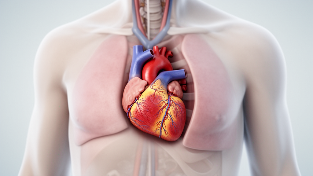
The clinical presentation initially raised suspicion of an acute myocardial infarction, but subsequent imaging revealed a rare and delayed complication: cardiac herniation into the left thoracic cavity. This case report, published in JA Clinical Reports, documents an exceptionally rare instance of cardiac herniation occurring in the chronic postoperative phase following minimally invasive thoracic surgery. The authors note that while cardiac herniation is a known potential complication of pericardial incision or resection, most reported cases develop intraoperatively or within the first 24 to 48 hours after surgery. Delayed-onset cases following minimally invasive procedures had not been previously reported in the medical literature.
The patient, who had a history of dyslipidemia, had undergone a video-assisted thoracoscopic extended thymectomy via a bilateral approach. No intraoperative pericardial injury was reported at the time of the initial surgery. Electrocardiography at a referring hospital showed ST-segment elevation in leads I, II, aVL, and V1–V4, a pattern typically associated with ST-elevation myocardial infarction. However, computed tomography of the chest demonstrated that the heart had herniated into the left thoracic cavity, providing an alternative explanation for her symptoms and electrocardiographic findings.
Further diagnostic evaluation using coronary angiography revealed significant stenosis in the distal segment of the left anterior descending artery, the second diagonal branch, and the obtuse marginal branch. The angiography also showed that the posterolateral branch was occluded. These areas of stenosis corresponded directly to the location of the hernia orifice, leading to a diagnosis of coronary artery compression secondary to cardiac herniation. Cardiac CT angiography confirmed these findings, showing stenosis at the same sites identified during the invasive angiography. The compression of the coronary arteries by the herniated heart tissue was presumed to be the cause of the acute myocardial infarction symptoms.
Contrast-enhanced computed tomography provided detailed visualization of the herniation, showing the left ventricle protruding from the mid-ventricular to the apical region into the left thoracic cavity.
Given the patient’s hemodynamic stability and the resolution of symptoms with conservative management, percutaneous coronary intervention was deferred. Instead, she was admitted for careful hemodynamic monitoring and initiated on pharmacotherapy with an oral coronary vasodilator. She was subsequently referred to a specialized institution for further evaluation and surgical planning. Chest radiography at the receiving hospital showed no apparent abnormalities, but the contrast-enhanced CT scan provided the critical diagnostic evidence of the cardiac herniation.
The patient underwent elective surgical repair of the cardiac hernia and coronary artery bypass grafting two months after the onset of symptoms. The surgical procedure involved hernia reduction and off-pump coronary artery bypass grafting using the left internal thoracic artery to the left anterior descending artery. The pericardial defect was repaired with a Gore-Tex patch. During the surgery, the heart was temporarily retracted to confirm cardiac compression at the hernia orifice. This maneuver required a temporary increase in the norepinephrine infusion rate to maintain blood pressure, but the infusion was successfully discontinued before the completion of the surgery.
Anesthetic management was carefully planned to address the potential circulatory changes associated with hernia reduction. The medical team anticipated that the release of cardiac restriction could lead to increased preload and an abrupt elevation of blood pressure, a phenomenon similar to that observed during the relief of cardiac tamponade. To prevent circulatory collapse due to anesthesia-induced afterload reduction, continuous administration of vasoconstrictors was planned from the induction of anesthesia. However, intraoperative monitoring revealed minimal changes in hemodynamic parameters during hernia reduction. The norepinephrine infusion rate was maintained at a low level, and no additional vasoactive agents were required, suggesting that the physiological changes accompanying herniation reduction were limited in this case.
The postoperative course was uneventful, and the patient was extubated on postoperative day zero. She was discharged on postoperative day seven without complications. The favorable outcome in this case of partial herniation may be attributed to the fact that the herniation resulted in only limited stenosis of the coronary artery without significant circulatory instability. The myocardial ischemia caused by coronary compression was reversible following coronary artery bypass and decompression. This case highlights the potential for a favorable clinical course even in cases of partial cardiac herniation, provided that the compression does not lead to severe circulatory compromise.
Cardiac herniation is classified into complete and partial types, with partial herniation generally considered to carry a poorer prognosis due to the higher risk of cardiac incarceration and myocardial ischemia. However, the present case demonstrates that a favorable outcome is possible. The precise mechanisms underlying the delayed onset of cardiac herniation six years after surgery remain unclear. The authors suggest that a small, unrecognized pericardial defect may have gradually enlarged over time, ultimately resulting in cardiac herniation. This hypothesis is plausible given the limited operative field and visualization inherent to thoracoscopic surgery.
The increasing number of minimally invasive procedures, including robot-assisted and thoracoscopic surgeries, due to their advantages in reducing postoperative pain and promoting early recovery, underscores the importance of recognizing potential late complications. This case serves as a reminder of the risk of delayed cardiac herniation in the chronic postoperative phase following minimally invasive thoracic surgery. Meticulous intraoperative inspection of the pericardium and prophylactic closure or reinforcement of even minor defects, when identified, may represent potential preventive measures. The case highlights the need for long-term follow-up and awareness of this rare but potentially fatal complication in patients who have undergone thoracic surgery involving pericardial incision or resection.
The clinical manifestations of cardiac herniation range widely from asymptomatic subjects to sudden cardiac death, depending on the presence and combination of cardiac displacement, torsion of the great vessels, associated inflow or outflow obstructions, and strangulation at the herniation orifice. The hemodynamic consequences are influenced by the location and size of the herniation defect. Right-sided cardiac herniation is more likely to cause reduced venous return and cardiac output due to torsion at the atrioventricular junction and bilateral ventricular outflow tracts. In contrast, left-sided cardiac herniation tends to result in myocardial and coronary artery compression at the herniation orifice, as observed in this case.
While cardiac herniation is a rare event, its occurrence six years after thoracoscopic thymectomy is exceptionally rare and has not been previously reported. The case underscores the importance of considering cardiac herniation in the differential diagnosis of patients presenting with chest symptoms or hemodynamic instability long after thoracic surgery.
The authors emphasize that cardiac herniation may occur even long after minimally invasive thoracic surgery and should be considered in patients presenting with chest symptoms or hemodynamic instability.
In conclusion, this case report documents an exceptionally rare instance of cardiac herniation manifesting six years after thoracoscopic thymectomy. The patient presented with symptoms mimicking an acute myocardial infarction, but imaging revealed the underlying mechanical cause. This case serves as a reminder of the potential risk of delayed cardiac herniation in the chronic postoperative phase following minimally invasive thoracic surgery and underscores the need for long-term awareness and follow-up in such patients.
Subject of Research: Delayed cardiac herniation as a rare complication of thoracoscopic thymectomy
Article Title: Cardiac herniation six years after thoracoscopic thymectomy: a case report
Article References: Ishizuka, Y., Hayashida, T., Yoshii, T., & Hasegawa-Moriyama, M. (2026). Cardiac herniation six years after thoracoscopic thymectomy: a case report. JA Clinical Reports, 12(1), Article 21. https://doi.org/10.1186/s40981-026-00856-6
Image Credits: AI Generated
DOI: 10.1186/s40981-026-00856-6
Keywords: cardiac herniation, thoracoscopic thymectomy, myasthenia gravis, coronary artery compression, pericardial defect, minimally invasive surgery, delayed complication, coronary artery bypass grafting, hemodynamic instability, chest pain, myocardial infarction, pericardial resection, thoracic surgery, case report, JA Clinical Reports, postoperative complication, coronary stenosis, hernia reduction, Gore-Tex patch, anesthetic management
News Source: Ophelia Keating. (November 21, 2026). Heart herniates six years after minimally invasive thymectomy. Scienmag.