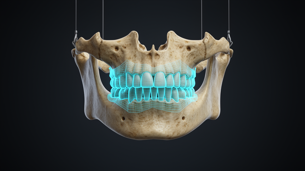
A retrospective proof-of-concept study has demonstrated the technical feasibility of using a deep learning architecture to automatically segment mandibular bone invasion in preoperative computed tomography examinations of patients with oral squamous cell carcinoma. Identifying the precise boundary of tumor invasion into the lower jawbone is essential for virtual surgical planning, yet manual delineation on CT scans remains challenging and time-consuming for clinical teams.
To address this clinical challenge, researchers investigated whether automated three-dimensional segmentation could delineate both the mandible and radiologically apparent bone invasion, while exploring centerline-based evaluations of longitudinal tumor extent for computer-aided design and computer-aided manufacturing planning. The study evaluated preoperative CT examinations from 50 patients who had histopathologically confirmed mandibular invasion by oral squamous cell carcinoma.
The cohort was divided into two distinct groups, with 30 CT examinations allocated for model development and the remaining 20 examinations reserved for independent testing. Model training employed a three-dimensional MedNeXt architecture implemented within the nnU-Net framework, utilizing expert-reviewed CT reference segmentations as ground truth to train the automated network.
Model performance was evaluated across multiple standardized metrics, including the Dice similarity coefficient, precision, recall, average symmetric surface distance, and the 95th-percentile Hausdorff distance. In the independent holdout testing cohort, researchers specifically analyzed spatial localization, longitudinal invasion-span differences, and signed centerline-projected endpoint errors to determine anatomical fidelity.
The deep learning model achieved strong segmentation accuracy for the healthy mandibular bone structure, recording a mean mandibular Dice similarity coefficient of 0.965 with a 95 percent confidence interval ranging from 0.961 to 0.969 and a median of 0.967. Delineating the invasive tumor tissue proved more challenging, yielding a mean invasion Dice similarity coefficient of 0.553 with a 95 percent confidence interval between 0.425 and 0.660, accompanied by a median of 0.623.
For the detection of mandibular invasion, the network attained a mean precision of 0.742 and a mean recall of 0.483. Predicted tumor invasion overlapped the CT reference annotations in 18 out of the 20 test examinations, representing a 90 percent overlap rate, while 16 of the 20 cases achieved a Dice similarity coefficient of 0.20 or higher. Two cases were completely missed by the automated algorithm.
Among the 18 evaluable cases showing spatial overlap, the median absolute invasion-span difference was 7.01 millimeters, with an interquartile range extending from 5.08 to 13.00 millimeters. Furthermore, the evaluation revealed a median symmetric endpoint distance of 5.88 millimeters, with an interquartile range spanning 3.19 to 9.16 millimeters along the centerline projection.
The authors concluded that automated CT segmentation of the mandible and radiologically apparent bone invasion is technically feasible and may assist in transferring radiological tumor extent into virtual resection workflows. However, the researchers emphasized that larger multicenter studies and prospective clinical validations remain necessary to definitively confirm planning accuracy and workflow efficiency.
Subject of Research: Deep learning segmentation of mandibular bone invasion in oral squamous cell carcinoma
Article Title: Deep learning–based CT segmentation of mandibular bone invasion in oral squamous cell carcinoma
Article References: Steybe, D., Fiederling, E., Poxleitner, P., Otto, S., Obermeier, K. T., Sabel, B., Rau, S., Reisert, M., & Russe, M. F. (2026). Deep learning–based CT segmentation of mandibular bone invasion in oral squamous cell carcinoma. BMC Oral Health, 26(1), Article 2362. https://doi.org/10.1186/s12903-026-10471-7
Image Credits: AI Generated
DOI: 10.1186/s12903-026-10471-7
Keywords: oral squamous cell carcinoma, mandibular invasion, deep learning, computed tomography, medical image segmentation, virtual surgical planning, CAD/CAM, MedNeXt, nnU-Net, Dice similarity coefficient, bone segmentation, reconstructive surgery, head and neck oncology, surgical planning, precision oncology, proof of concept
News Source: Blake Davidson. (November 21, 2026). Deep Learning CT Segmentation of Mandibular Bone Invasion Demonstrates Technical Feasibility. Scienmag.