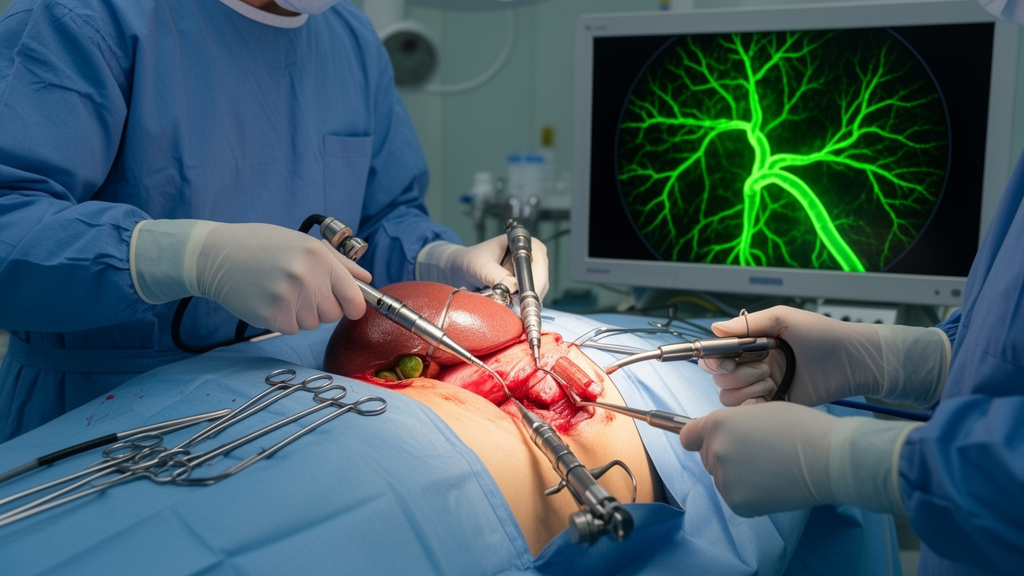
Laparoscopic cholecystectomy in patients with situs inversus totalis presents a unique technical challenge due to the complete mirror-image transposition of thoracoabdominal organs. A recent case report published in Innovative Surgical Trends describes the successful use of indocyanine green (ICG) fluorescence cholangiography to guide intraoperative bile duct visualization during this procedure. The study, led by Kyung Young Kim and colleagues, highlights how advanced imaging techniques can mitigate the risks associated with reversed anatomical orientation in this rare congenital condition. The authors demonstrate that ICG fluorescence serves as a valuable adjunct to standard laparoscopic techniques, enhancing the surgeon’s ability to identify critical biliary structures in real time.
Situs inversus totalis is a rare congenital condition characterized by the complete reversal of the normal position of the internal organs. In affected individuals, the liver and gallbladder are located in the left upper quadrant rather than the right, which complicates both diagnostic imaging and surgical intervention. The authors note that while the incidence of cholelithiasis and cholecystitis in patients with situs inversus totalis is similar to that of the general population, the altered anatomy can lead to significant diagnostic and intraoperative uncertainty. This uncertainty increases the risk of complications, particularly bile duct injury, which is a significant source of postoperative morbidity in cholecystectomy.
The primary objective of the reported procedure was to safely perform a laparoscopic cholecystectomy in a patient with situs inversus totalis by utilizing intraoperative indocyanine green fluorescence cholangiography. The surgical team aimed to use this technology to guide the identification of biliary structures and facilitate real-time biliary mapping. By employing this method, the surgeons sought to overcome the spatial disorientation caused by the reversed anatomy and ensure the safe dissection of the hepatocystic triangle.
Preoperative planning involved a comprehensive imaging protocol to delineate the patient’s hepatobiliary anatomy and exclude any biliary anomalies or duct dilatation. The team utilized ultrasound, computed tomography, and magnetic resonance imaging to map the reversed organ positions. The preoperative CT scan confirmed situs inversus totalis, revealing the liver and gallbladder in the left upper quadrant. This detailed preoperative assessment was crucial for planning the surgical approach and ensuring that the anatomical variations were fully understood before the operation began.
The surgical technique involved a specific modification of the standard laparoscopic port placement to accommodate the reversed anatomy. Under general anesthesia, the patient was positioned supine, and pneumoperitoneum was achieved via a Veress needle inserted at reverse Palmer’s point on the patient’s right side. Four trocars were placed in a mirror-image configuration, including a 5 mm periumbilical optical port, a 10 mm epigastric working port, and two 5 mm ports along the left mid-clavicular and anterior axillary lines. This configuration allowed the primary surgeon to stand on the patient’s right side and perform the dissection with the non-dominant hand, while the assistant stood on the left.
Indocyanine green, a low-toxicity fluorescent agent that is excreted exclusively via the biliary system, was administered intravenously one hour before the surgery. The dose used was 0.5 mL of a 2.5 mg/mL solution. ICG enables dynamic visualization of biliary anatomy under near-infrared imaging, providing a clear distinction between bile ducts and surrounding tissues. The authors note that studies have shown ICG to be a safe and valid alternative to standard intraoperative cholangiography, which typically uses contrast dye and fluoroscopy.
During the procedure, the surgical team alternated between standard white-light imaging and near-infrared imaging using the Stryker 1488 AIM Laparoscopy Tower System. Diagnostic laparoscopy first confirmed the situs inversus totalis, with the liver and gallbladder clearly visible in the left hemi-abdomen. The gallbladder was identified and retracted cephalad to expose Calot’s triangle. The ICG fluorescence provided clear, real-time visualization of the cystic duct and common bile duct, allowing the surgeons to distinguish these structures from the surrounding tissue with high precision. This enhanced visualization facilitated the safe dissection of the hepatocystic triangle and the establishment of the critical view of safety.
The results of the case report indicate that the laparoscopic cholecystectomy was successfully completed without any intraoperative or postoperative complications. The ICG fluorescence cholangiography played a key role in aiding the dissection of Calot’s triangle by providing a clear map of the biliary anatomy. The cystic duct and artery were clipped and divided without difficulty, and the gallbladder was dissected from the liver bed and extracted through the epigastric port. The total operative time was within the expected range for an elective laparoscopic cholecystectomy, suggesting that the use of ICG did not significantly prolong the procedure.
The patient recovered uneventfully and was discharged on postoperative day one, further supporting the safety and efficacy of the approach. The authors emphasize that the use of ICG fluorescence cholangiography helped mitigate the risks associated with anatomical variation in situs inversus totalis. By providing a clear view of the biliary structures, the technology allowed the surgical team to maintain a high level of spatial awareness and adapt to the reversed anatomical landmarks. This case demonstrates that advanced imaging tools can be effectively integrated into standard surgical workflows to improve outcomes in complex cases.
The study concludes that laparoscopic cholecystectomy in patients with situs inversus totalis is rarely described in the literature and remains technically demanding due to the need for reorientation of standard visuomotor coordination. The authors suggest that intraoperative ICG fluorescence cholangiography offers enhanced anatomical orientation and real-time visualization, which may reduce the risk of bile duct injury. This approach serves as a safe and effective adjunct to standard laparoscopic techniques, particularly in anatomically complex cases where traditional landmarks are reversed.
In conclusion, the use of indocyanine green fluorescence cholangiography during laparoscopic cholecystectomy in a patient with situs inversus totalis represents a successful application of advanced imaging technology in a complex surgical setting. The procedure was completed without complications, and the ICG imaging provided critical real-time visualization of the biliary anatomy.
Subject of Research: Use of ICG fluorescence cholangiography for bile duct visualization during laparoscopic cholecystectomy in situs inversus totalis
Article Title: The use of indocyanine green (ICG) fluorescence cholangiography for intraoperative bile duct visualization during laparoscopic cholecystectomy in situs inversus totalis
Article References: Kim, K. Y., Mikhail, A., Chadi, S., & Kazazian, K. (2025). The use of indocyanine green (ICG) fluorescence cholangiography for intraoperative bile duct visualization during laparoscopic cholecystectomy in situs inversus totalis. Innovative Surgical Trends, 1(1), Article 4. https://doi.org/10.1007/s44414-025-00004-0
Image Credits: AI Generated
DOI: 10.1007/s44414-025-00004-0
Keywords: indocyanine green, fluorescence cholangiography, laparoscopic cholecystectomy, situs inversus totalis, bile duct visualization, near-infrared imaging, biliary anatomy, critical view of safety, intraoperative imaging, surgical anatomy, congenital anomalies, minimally invasive surgery, hepatobiliary surgery, port configuration, real-time visualization, surgical safety, anatomical variation, case report, biliary mapping, operative complexity
News Source: Ophelia Keating. (November 20, 2026). Fluorescent dye aids bile duct visualization in rare anatomy case. Scienmag.