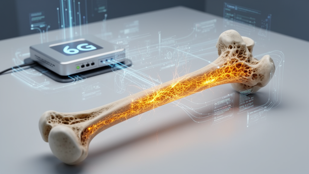
A computational framework combining artificial intelligence, dynamic digital twin modeling, and simulated sixth-generation wireless networking has demonstrated high analytical accuracy and ultra-low latency in preclinical tests designed to detect skeletal tissue injury in bone cancer patients. Researchers developed the system, designated AI-6G-DT-Sys, to address persistent clinical challenges where conventional diagnostic modalities often fail to identify early microstructural deterioration, leading to diagnostic delays, invasive biopsy procedures, and escalated clinical costs. The investigative architecture was evaluated across secondary, publicly accessible medical imaging databases alongside simulated telecommunication network profiles, establishing technical feasibility for continuous physical-to-digital state synchronization without relying on invasive physical hardware deployments.
The study specifically targets skeletal tissue injury, defined strictly as structural abnormalities within bone tissue, rather than treating tumors, necrosis, microfractures, or broad tissue damage as interchangeable clinical concepts. In bone cancer pathology, traditional diagnostic protocols frequently exhibit insufficient sensitivity to capture initial microstructural variations before evident symptoms or macroscopic fractures occur. Compounding these diagnostic limitations are technical bottlenecks associated with fusing heterogeneous imaging modalities, safeguarding sensitive medical data transmissions, and maintaining synchronous computational models of patient physiology in near-real-time environments. By integrating multimodal deep learning with ultra-reliable low-latency communication profiles, the research team designed a unified pipeline capable of tracking dynamic disease trajectories while managing distributed network workloads.
Rather than asserting a fully realized patient-specific replica from individual subjects, the investigators formulated AI-6G-DT-Sys as a population-informed digital twin simulation framework. This architectural decision was necessitated by the characteristics of the underlying research data, which comprised three independent, unlinked secondary datasets: the Osteosarcoma Tumor Assessment dataset for histopathology, the Spine-Mets-CT-SEG dataset containing computed tomography volumes from patients with metastatic spine disease, and the Bone Cancer MRI Dataset from Kaggle. Because these secondary repositories lacked subject-level correspondence across modalities, the framework did not reconstruct individualized patient anatomy from matched observations. Instead, each imaging modality was processed to learn population-level structural, radiological, and cellular representations that were subsequently integrated into a shared latent space via label-consistent statistical normalization.
The technical architecture processes clinical imaging across macro, meso, and micro scales by deploying dedicated, modality-specific feature encoders. Computed tomography scans, standardized into Hounsfield Units to quantify tissue radiodensity against linear attenuation baselines of water and air, are processed through three-dimensional convolutional neural networks or Vision Transformers to map skeletal geometry, bone density variations, and lesion margins. Magnetic resonance imaging volumes are standardized through volumetric z-score normalization and analyzed using convolutional architectures to delineate marrow infiltration and soft-tissue margins. For microscopic evaluation, gigapixel whole-slide histopathology images undergo patch-based tiling into discrete 256-by-256 pixel arrays, color normalization via Reinhard or Macenko techniques to remove staining variability, and feature extraction via patch-level neural encoders to capture cellular necrosis and tumor viability.
To fuse these disparate feature representations without artificial patient pairing, the system projects modality embeddings into a unified latent manifold optimized through contrastive alignment objectives. The cross-modal alignment mechanism employs temperature-scaled similarity metrics to ensure that semantically comparable pathological states occupy adjacent regions within the feature space. Following alignment, an attention-based fusion module dynamically assigns diagnostic weights to each modality based on learned relevance parameters and feature confidence. This adaptive weighting enables the model to adjust focus when certain streams, such as real-time histopathological data, are unavailable or degraded, thereby preventing spurious correlations and maintaining robust joint inferences across macrostructural and cellular domains.
Communication demands and real-time state synchronization were simulated using the ns-3 network emulator configured with simulated 6G communication attributes, rather than live physical cellular transmissions. The simulated radio environment assumed a single base station serving twenty user equipments operating over a 28 GHz carrier frequency, 100 MHz bandwidth, and 30 dBm transmission power, employing proportional-fair scheduling and three-dimensional propagation channel models under UDP constant-bit-rate traffic. Under these standardized parameters, the end-to-end inference and synchronization pipeline achieved an average latency of 0.62 milliseconds, representing a 68.4 percent reduction compared to a baseline fifth-generation ultra-reliable low-latency communication profile. Furthermore, the framework demonstrated simulated energy savings of 41.7 percent at sensing nodes and 33.2 percent at edge servers compared to static allocation schemes.
Data governance and transaction logging were incorporated using a Practical Byzantine Fault Tolerance consensus protocol optimized for hospital edge deployments. Rather than routing large volumetric images or gigapixel histopathology slides through the distributed ledger, the blockchain layer handles lightweight metadata, feature hashes, state checkpoints, and access-control verifications asynchronously. In a ten-validator network configuration, the consensus mechanism achieved a steady throughput of approximately 1,250 transactions per second with an average finalization latency of 3.4 milliseconds, capped at 5 milliseconds under peak loads. Because ledger writes operate outside the critical inference pathway, cryptographic validation introduced no latency bottlenecks to the sub-millisecond diagnostic data transfer path.
In experimental evaluations using a controlled seventy-fifteen-fifteen train, validation, and test partition across the multimodal datasets, AI-6G-DT-Sys demonstrated substantial diagnostic improvements over baseline medical artificial intelligence models. The system converged to a classification accuracy of approximately 95.7 percent, outperforming baseline models by Yuan and colleagues, Rajak and colleagues, and Zhao and colleagues, which stabilized between 85 percent and 89 percent. Multi-task learning simultaneously optimized segmentation, classification, and prognostic risk scoring, yielding an Area Under the Receiver Operating Characteristic curve of approximately 0.96 and a Dice segmentation coefficient of 0.91 at epoch ninety. In ablation testing, multimodal feature fusion accounted for an estimated 55 to 60 percent of observed accuracy and Dice gains, while advanced neural modeling contributed 25 to 30 percent, and temporal digital twin state updates contributed the remaining 10 to 15 percent.
The system integrates an interactive clinical decision support interface designed to translate multi-task computational predictions into actionable visualizations. Clinicians can review three-dimensional skeletal heatmaps displaying cortical thinning and microfractures alongside coregistered histopathological reports indicating necrotic versus viable tumor cell ratios. To mitigate the danger of over-interpreting predictions derived from corrupted inputs, the dashboard incorporates uncertainty-aware quality gates. When input computed tomography or magnetic resonance scans exhibit severe acquisition artifacts or missing slices, the platform issues explicit low-confidence alerts and suppresses automated recommendations, directing practitioners to examine raw diagnostic scans rather than accepting synthetic forecasts as confirmed clinical outcomes.
Despite promising simulation and offline computational metrics, the researchers explicitly emphasized that AI-6G-DT-Sys remains an unvalidated preclinical research prototype not yet suitable for direct patient diagnosis or clinical deployment. The evaluation lacked true patient-level linkage across imaging modalities, relying on population-level statistical harmonization rather than anatomically matched, longitudinal clinical datasets acquired from identical patients over time. Furthermore, telecommunication evaluations were confined to simulator software without validation on physical 6G hardware, which remains unstandardized and undeployed. Future work must focus on assembling longitudinally matched multi-institutional cohorts, prospective clinical trials, clinician-in-the-loop assessments, and physical hardware testbed validations to establish clinical safety, regulatory compliance, and operational reliability in hospital environments.
Optimization of the multimodal architecture relied on the Adam optimizer configured with an initial learning rate of 0.0001, decay parameters of 0.9 and 0.999, and an L2 regularization coefficient of 0.00001 to stabilize convergence across heterogeneous imaging streams. Memory constraints associated with volumetric CT and MRI scans dictated a batch size of 16, whereas histopathology patch training utilized a batch size of 32 backed by gradient accumulation across 120 training epochs per encoder. Most networks converged within 90 to 105 epochs. The multimodal fusion network incorporated four fully connected layers dimensioned at 512, 256, 128, and 64 nodes, coupled to an eight-head cross-modal attention block with a key-query-value dimension of 64 and a temperature scaling factor of 0.7 to enforce sharp modality differentiation.
To evaluate model stability against real-world data corruption, the investigators simulated preprocessing perturbations across input streams. Introducing boundary segmentation noise of plus or minus 5 to 15 percent into CT lesion masks caused limited feature drift, resulting in accuracy drops between 1.8 and 4.9 percent and a Dice reduction of 0.03 at maximum perturbation. For histopathology slides, varying patch tiling overlaps between zero and 30 percent alongside simulated tissue dropouts produced a peak accuracy decrease of 3.6 percent, preserved by multi-instance aggregation. When multi-source preprocessing noise occurred simultaneously, uncertainty-aware cross-modal attention restricted overall diagnostic degradation to below 6.2 percent.
Network stress testing further revealed structural resilience under unstable transmission conditions. At extended network latencies of 100 milliseconds, the framework sustained an F1-score of 83.1 percent, compared to 72.9 percent and 65.0 percent for baseline models. Implementing semantic-aware transmission restricted packet losses between 3 and 5 percent to minor latency jitter, keeping classification drift under 2.4 percent through temporal twin smoothing. Under intermittent disconnects, an edge-autonomous fallback mode executed local inference from the latest synchronized state, reducing edge computational workloads by 38 percent with a minor 3 to 4 percent accuracy penalty. In component ablation, removing multimodal fusion decreased the overall F1-score from 93.83 percent to 91.20 percent, while omitting the digital twin component reduced performance to 93.29 percent.
Subject of Research: 6G-oriented digital twin and artificial intelligence simulation for skeletal tissue injury detection
Article Title: A 6 G-Oriented simulation framework for ultra-reliable low-latency communication in real-time skeletal tissue injury detection
Article References: Nagappan, V. K., Sivamani, S., Kalai Selvi, K., & Prasanna Bharathi, S. (2026). A 6 G-Oriented simulation framework for ultra-reliable low-latency communication in real-time skeletal tissue injury detection. Discover Computing, 29(1), Article 916. https://doi.org/10.1007/s10791-026-10618-8
Image Credits: AI Generated
DOI: 10.1007/s10791-026-10618-8
Keywords: artificial intelligence, digital twin, 6G simulation, skeletal tissue injury, bone cancer, multimodal deep learning, computed tomography, magnetic resonance imaging, histopathology, ultra-reliable low-latency communication, edge computing, blockchain, practical byzantine fault tolerance, clinical decision support, ns-3 simulator, image segmentation
News Source: Blake Davidson. (November 20, 2026). Simulated 6G Digital Twin Framework Evaluated for Bone Tissue Injury Detection. Scienmag.