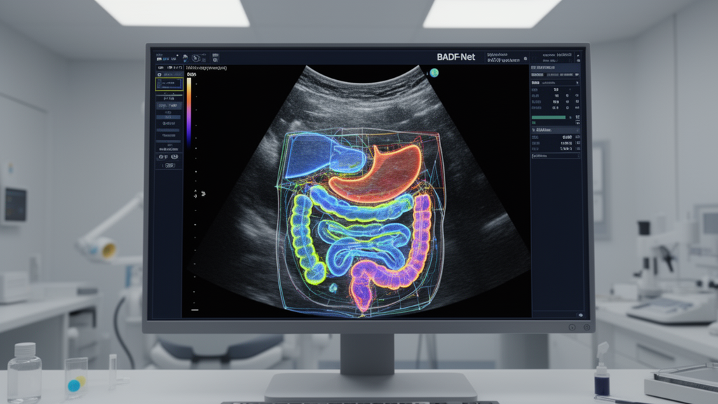
In an original research study published in Frontiers in Medicine, an international team of researchers demonstrated that their proposed BADF-Net deep learning architecture outperformed eleven baseline models in segmenting multiple anatomical structures from abdominal ultrasound scans. Across a multi-anatomical kidney and spleen ultrasound dataset, the neural network achieved an overall Dice score of 0.8174 alongside a Pixel Accuracy of 0.9703 and a Mean Pixel Accuracy of 0.8521. The evaluation demonstrated that the model produced the lowest 95th percentile Hausdorff distance, recorded at 11.49, marking a substantial reduction in boundary error compared with existing baseline approaches.
The investigation addressed longstanding engineering obstacles in automated ultrasound analysis, where imaging artifacts consistently compromise deep learning performance. Specifically, the authors highlighted that speckle noise, low acoustic contrast, and weak anatomical boundaries impede conventional automated segmentation models from reliably delineating internal organ borders. The researchers emphasized that consistent quantitative measurements derived from automated multi-anatomical segmentation are critical for clinical decision-making and longitudinal disease monitoring.
To resolve boundary ambiguity, the authors designed BADF-Net around a squeeze-and-excitation recalibrated ResNet-34 encoder. At every sequential stage of this encoder, the network integrates a Boundary-Aware Dynamic Attention module paired with Boundary Confidence Estimation.
Building upon the recalibrated encoder representations, the architecture introduces a Cross-Scale Boundary Fusion module. This module adaptively combines multi-scale boundary cues extracted across different network depths into a unified, shared guidance feature representation. In the subsequent phase, BADF-Net routes these combined cues into a specialized boundary-guided decoder that incorporates attention-gated skip fusion mechanisms. Through these interconnected skip connections, the decoder selectively merges localized low-level boundary indicators with high-level spatial abstractions, enhancing structural boundary localization and tissue discrimination across multiple adjacent organs.
When evaluated against expert radiologist annotations on the multi-anatomical kidney and spleen dataset, the architecture achieved its most pronounced performance improvements in challenging, low-contrast internal regions, specifically within the kidney cortex and medulla. Conversely, agreement with the expert annotations reached its highest numerical peak on geometrically well-defined organs, recording a score of 0.9786 for spleen segmentation.
Beyond geometric precision metrics, the experimental evaluation showed that BADF-Net processed individual ultrasound images in 0.016 seconds, meeting requirements for real-time computational integration into clinical workflows. The investigators noted that this processing speed, coupled with consistent automated boundary extraction, allows the system to operate as a reliable second-reader tool in high-volume hospital environments. In this clinical support capacity, the computational framework aims to reduce operator dependency and mitigate inter-observer variability during routine ultrasound interpretation without introducing latency into daily diagnostic routines.
The collaborative research was conducted by investigators across multiple academic institutions, including Pabna University of Science and Technology, Kocaeli University, Green University of Bangladesh, United International University, Malardalens universitet, and Brandon University. In their concluding discussion, the authors affirmed that dynamic boundary attention, confidence-weighted feature refinement, and channel recalibration jointly resolve critical ultrasound segmentation obstacles with direct clinical applicability. To facilitate replication and further technological development, the research team made their complete experimental code publicly available through an open-access GitHub repository.
Subject of Research: Multi-anatomical structure segmentation in abdominal ultrasound imaging
Article Title: BADF-Net: a boundary-aware dynamic fusion network for robust multi-anatomical structure segmentation in abdominal ultrasound
Article References: Rashed, M., Jamil, M., Hossain, M. I., Kavak, A., Hassan, R., Ohidujjaman, O., Hosain, M. S., Çakır, Ö., Fotouhi, H., & Srivastava, G. (2026). BADF-Net: a boundary-aware dynamic fusion network for robust multi-anatomical structure segmentation in abdominal ultrasound. Frontiers in Medicine, 13, 1998681. https://doi.org/10.3389/fmed.2026.1998681
Image Credits: AI Generated
DOI: 10.3389/fmed.2026.1998681
Keywords: BADF-Net, abdominal ultrasound, anatomical segmentation, kidney segmentation, spleen segmentation, kidney cortex, kidney medulla, boundary-aware dynamic attention, boundary confidence estimation, cross-scale boundary fusion, ResNet-34 encoder, squeeze-and-excitation, attention-gated skip fusion, Hausdorff distance, Dice score, pixel accuracy, real-time image processing, clinical decision support
News Source: Ophelia Keating. (November 20, 2026). BADF-Net Outperforms Eleven Baseline Models in Segmenting Abdominal Ultrasound Structures. Scienmag.