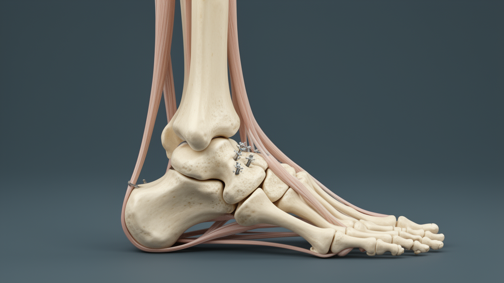
A novel anterior talar osteotomy provides effective structural correction and restores physical mobility in patients suffering from equinus deformity accompanied by posterior tilt of the talus trochlea. Orthopaedic researchers evaluated mid- and long-term clinical and radiological records to assess how well this joint-sparing intervention resolved complex ankle deformities. Their findings reveal substantial improvements in structural alignment and functional capacity over an extended observational timeframe.
Equinus deformity frequently presents with an abnormal posterior tilt of the talus trochlea, creating substantial challenges for conventional orthopaedic management. Standard soft-tissue interventions, including isolated Achilles tendon lengthening, often prove insufficient for achieving adequate anatomical correction and can induce tibiotalar joint incongruence. Conversely, definitive salvage options like triple arthrodesis permanently sacrifice articular motion, making an effective joint-preserving surgical alternative particularly desirable for active individuals.
To investigate the procedural efficacy of anterior talar osteotomy, the surgical team conducted a Level IV case series analyzing twelve patients with a mean age of 26.83 ± 8.83 years. In addition to the surgical cohort, investigators recruited fifty healthy individuals to establish baseline anatomical measurements. These healthy control subjects enabled precise morphological comparisons of the talar trochlea and confirmed the presence of pathological posterior tilt in the affected patient cohort.
Preoperative radiographic and computed tomography assessments confirmed severe baseline anatomical deviations among the twelve surgical candidates compared with the control cohort. Preoperative X-ray evaluation demonstrated an average talus trochlea–neck angle of 53.58 ± 9.93 degrees alongside a talar neck depth of 0.05 ± 0.04. Preoperative computed tomography scans corroborated these structural measurements, registering an average talus trochlea–neck angle of 51.89 ± 9.34 degrees.
The anatomical parameters recorded in the surgical cohort contrasted markedly with those measured across the fifty healthy control subjects. Within the healthy control group, the mean talus trochlea–neck angle on X-ray measured 30.58 ± 3.13 degrees, the talar neck depth averaged 0.30 ± 0.04, and the computed tomography angle averaged 25.57 ± 2.00 degrees. These baseline differences established clear quantitative diagnostic markers characterizing the patient group’s underlying morphological pathology.
Postoperative imaging obtained over an average follow-up duration of 80.92 ± 18.36 months demonstrated meaningful anatomical correction following the anterior osteotomy. The mean talus trochlea–neck angle on X-ray improved significantly to 38.00 ± 4.20 degrees, while the talar neck depth increased to 0.10 ± 0.05. These objective radiological changes confirmed that the surgical technique successfully realigned the bony architecture toward normal physiological values.
Patient-reported functional outcomes reflected substantial daily improvements, as measured by standardized scoring instruments throughout the longitudinal follow-up period. The Foot and Ankle Disability Index activity score rose dramatically from a preoperative baseline of 43.67 ± 12.23 to 91.92 ± 4.89 postoperatively. Concurrently, the Foot and Ankle Disability Index sports score improved from 6.58 ± 3.55 prior to surgery to 23.92 ± 3.65 at final follow-up.
Clinical evaluations documented an overall good and excellent outcome rate of 75 percent across the patient series. Crucially from a surgical safety perspective, all twelve talar osteotomies achieved complete bone union, and no cases of avascular necrosis of the talus were detected during the extended follow-up period. The absence of osteonecrosis confirms the biological viability and vascular safety of the osteotomy technique.
The study authors concluded that anterior talar osteotomy offers a viable, joint-sparing therapeutic avenue for correcting equinus deformity associated with posterior trochlear tilt. By maintaining tibiotalar joint congruence and preserving subtalar joint function without joint-sacrificing fusion, the researchers noted that this procedure represents an advantageous intervention particularly suited for young, physically active patients seeking long-term functional recovery.
Subject of Research: Clinical and radiological outcomes of anterior talar osteotomy for equinus deformity
Article Title: A novel anterior talar osteotomy for equinus deformity with posterior tilt of the talus trochlea: the mid- and long-term outcomes
Article References: Xu, Y., Yang, C., Guo, C., Zhu, Y., Yang, J., Xu, X., & Li, X. (2026). A novel anterior talar osteotomy for equinus deformity with posterior tilt of the talus trochlea: the mid- and long-term outcomes. Journal of Orthopaedic Surgery and Research. https://doi.org/10.1186/s13018-026-07338-6
Image Credits: AI Generated
DOI: 10.1186/s13018-026-07338-6
Keywords: anterior talar osteotomy, equinus deformity, talus trochlea, posterior tilt, tibiotalar joint congruence, subtalar joint preservation, Foot and Ankle Disability Index, talar neck depth, trochlea-neck angle, joint-sparing surgery, avascular necrosis, orthopaedic surgery, bone union, case series, computed tomography, radiographic imaging
News Source: Ophelia Keating. (November 20, 2026). Anterior Talar Osteotomy Corrects Equinus Deformity While Preserving Joint Congruence. Scienmag.