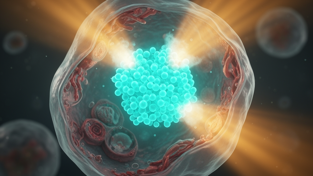
A research team led by the University of Osaka in Japan, in collaboration with the University of British Columbia in Canada, has synthesized an advanced polymer material capable of performing both cellular imaging and light-activated cell destruction within a single molecular architecture. Detailed in an experimental study published in Chem & Bio Engineering, the innovation addresses longstanding hurdles in nanotheranostics, an emerging biomedical discipline focused on integrating diagnostic visualization and therapeutic capabilities into unified nanoscale systems. By formulating the newly created polymer into specialized nanoparticles termed polymer dots, the scientists demonstrated that a singular synthetic platform can emit stable fluorescence for tracking cells while simultaneously generating cytotoxic reactive oxygen upon targeted light exposure.
Developing functional nanotheranostic agents has historically proven exceptionally difficult because diagnostic imaging and therapeutic interventions impose conflicting physical and chemical requirements on a single substrate. Conventional approaches often bypass this conflict by chemically incorporating a secondary, standalone light-activated compound known as a photosensitizer alongside an imaging agent. In contrast, the Osaka and British Columbia team engineered their polymer to natively combine both bioimaging and photodynamic capabilities, eliminating the reliance on a distinct photosensitizer and potentially streamlining future manufacturing and design workflows for light-based medical interventions.
Central to the diagnostic function of the material is an optical mechanism known as thermally activated delayed fluorescence, or TADF. By harnessing TADF, the polymer dots emit an orange fluorescence with an emission lifetime that persists substantially longer than the natural, short-lived autofluorescent glow characteristic of living cells and tissues.
The unique photophysical behavior and stability of the material are governed by specialized structural and electronic mechanisms. The polymer relies partially on through-space charge transfer, an unconventional electronic process wherein spatial interactions take place between molecular groups packed closely together through three-dimensional space rather than across the covalent chemical bonds connecting the backbone. Furthermore, the macromolecular architecture demonstrates conformational adaptability, giving the molecular framework the structural flexibility to adopt diverse spatial arrangements without sacrificing its key functional characteristics. Working in unison, through-space charge transfer and conformational flexibility prevent the self-quenching and emission degradation that normally arise when luminescent molecules are densely packed inside compact nanoparticles.
Maintaining optical and functional performance in aqueous conditions represents another critical technical bottleneck, as water mimics physiological environments where densely grouped fluorophores commonly lose their luminescence. Lead author Nae Aota emphasized that laboratory tests proved the polymer dots sustained their characteristic orange emission and long-lived delayed fluorescence in water, even when formulated with a high proportion of light-emitting units.
Biological testing in cell cultures confirmed the functional efficacy of the polymer dots inside living biological environments. Once introduced to cultured cells, the nanoparticles underwent cellular uptake and concentrated predominantly within lysosomes, specialized intracellular organelles tasked with degrading and recycling cellular waste. Inside these compartments, the intense orange fluorescence enabled clear optical visualization of cellular structures. When the researchers subsequently irradiated the internalized material under suitable light conditions, the polymer dots generated singlet oxygen, an aggressive, reactive form of oxygen that damages targeted structures and establishes the foundational mechanism for photodynamic therapy aimed at inducing cancer cell death.
Senior author Youhei Takeda and the international research group highlighted that the successful development of this conformationally adaptive TADF polymer underscores the productive synergy of cross-border partnerships, supported in part by the JST-ASPIRE program. While current investigations remain situated at the experimental cellular stage, the researchers noted that their dual-functioning single-material architecture establishes an important empirical foundation for next-generation multifunctional nanomaterials, offering a versatile blueprint for future studies targeting cancer and other complex pathologies requiring synchronized imaging and light-activated treatment.
The research was conducted at the University of Osaka in partnership with the University of British Columbia, supported by the JST-ASPIRE program. As one of Japan’s original imperial universities, the University of Osaka maintains a strategic mandate as a Designated National University Corporation to integrate fundamental science into innovations for human welfare and sustainable development.
The findings are documented in the journal Chem & Bio Engineering (DOI: 10.1021/cbe.6c00141). By demonstrating that a single molecular framework can simultaneously handle imaging and light-activated therapeutic tasks, the study establishes an empirical foundation for future nanotheranostic research. While laboratory results confirm that the polymer dots accumulate in cellular lysosomes and exert photodynamic effects upon irradiation, the researchers note these findings remain in the experimental stage.
Subject of Research: Multifunctional TADF polymer dots for cellular bioimaging and photodynamic therapy
Article Title: One material, two functions: Bioimaging and triggering cell death with light
Article References: One material, two functions: Bioimaging and triggering cell death with light. Original publication (Bibliographic details unavailable.)
Image Credits: AI Generated
DOI: Not provided
Keywords: nanotheranostics, polymer dots, thermally activated delayed fluorescence, TADF, bioimaging, photodynamic therapy, singlet oxygen, through-space charge transfer, conformational adaptability, lysosomes, fluorescence imaging, cancer cell death, luminescent materials, nanoparticles, Youhei Takeda, Zachary M Hudson, Nae Aota
News Source: Neil Sanderson. (November 19, 2026). Dual-Function Polymer Dots Enable Fluorescent Cell Imaging and Light-Activated Therapy. Scienmag.