A multicenter neuroimaging investigation utilizing data from the Adolescent Brain Cognitive Development study revealed that youth diagnosed with an autism spectrum disorder exhibit a significant reduction in left amygdala functional activation when processing emotional faces compared to neutral faces, alongside a distinctive positive relationship between local neuron cell body density and task-evoked neural responses. Published in the Journal of Neurodevelopmental Disorders, the study evaluated both functional magnetic resonance imaging and advanced diffusion weighted imaging to examine whether cellular architecture in specific brain regions accounts for long-standing inconsistencies in autism-related functional imaging literature.
To investigate these neural mechanisms, the research team analyzed longitudinal datasets drawn from release 6.0 of the Adolescent Brain Cognitive Development study, a nationwide research initiative that initially recruited 11,873 children aged 9 to 10 across 21 active research sites in the United States. The current analysis identified 149 participants with a parent-reported autism spectrum diagnosis who also scored above the clinical threshold of 60 on the brief Social Responsiveness Scale, ensuring diagnostic validity in the absence of in-person clinical re-evaluations. For comparative baseline analysis, the researchers identified 10,146 individuals who had no reported diagnosis of autism. Data were gathered across four sequential collection waves spanning six years, incorporating scans obtained at baseline, two-year follow-up, four-year follow-up, and six-year follow-up intervals for participants between 9 and 16 years of age.
Participant selection adhered to rigorous exclusion criteria designed to remove confounding neurodevelopmental, medical, and technical variables. Children were excluded if they possessed uncorrectable sensory impairments such as legal blindness, lacked fluency in English, or had psychiatric conditions that prevented regular classroom attendance. Additional medical exclusion parameters ruled out participants with prior neurological illness, brain tumors, head trauma involving loss of consciousness for more than thirty minutes, or severe perinatal complications including gestational age under 28 weeks, birth weight below 1.2 kilograms, or neonatal hospitalization exceeding one month. All structural, diffusion, and functional scans underwent automated and radiologist-reviewed quality control pipelines to eliminate data corrupted by head motion, magnetic field artifacts, or file errors, and participants who scored below 60 percent accuracy on the experimental cognitive paradigm were removed from subsequent evaluations.
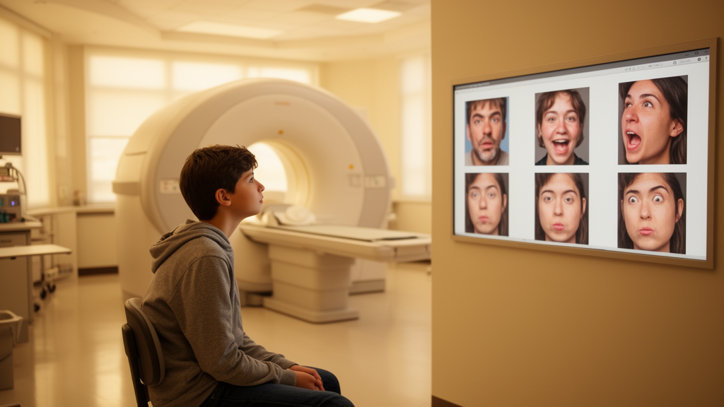
Functional responses to socio-emotional stimuli were measured during functional magnetic resonance imaging acquisition using the emotional n-back task across 160 experimental trials. The task presented children with visual stimuli consisting of 24 unique happy faces, 24 unique fearful faces, and 24 unique neutral faces, totaling 72 distinct facial photographs presented one at a time. The paradigm was administered in four task blocks across five-minute scanning runs and featured two operational variants: a zero-back condition requiring button presses to identify immediate face repeats and a two-back condition requiring working memory recognition of stimuli shown two iterations earlier. Each trial displayed an emotional face for two seconds followed by a 500-millisecond fixation cross, providing task-driven engagement of occipital, temporal, frontal, and limbic structures implicated in human facial processing.
To characterize underlying cellular morphology in vivo, the investigators implemented restriction spectrum imaging derived from diffusion weighted imaging acquired with 96 noncollinear gradient directions across multiple diffusion weightings, including b-values of 500, 1000, 2000, and 3000 seconds per square millimeter alongside unweighted frames. Restriction spectrum imaging separates intracellular fluid diffusion from extracellular hindered compartments, isolating discrete histological elements. Restricted normalized isotropic diffusion was measured to estimate the packing density of spherical neuron cell bodies, where water molecules diffuse evenly in all directions. Conversely, restricted normalized directional diffusion was quantified to characterize anisotropic water movement along cylindrical neurite projections, including dendrites and axons. These parameters overcome limitations of conventional diffusion tensor imaging by remaining robust in tissue voxels containing complex crossing fibers.
Processing of functional neuroimaging data used general linear models implemented in Analysis of Functional NeuroImages software to model hemodynamic responses convolved with a two-parameter gamma basis function. Individual functional contrasts isolated activation specifically evoked when viewing emotional expressions compared to neutral expressions, producing a subject-level metric termed estimated functional activity while processing emotional versus neutral faces. High-resolution T1-weighted and T2-weighted structural scans were corrected for gradient nonlinearity and spatial intensity variations, registered, and parcellated according to the Desikan-Killiany brain atlas via FreeSurfer software to extract mean diffusion and functional values within bilateral regions of interest comprising the amygdala, fusiform cortex, and inferior frontal cortex.
Statistical modeling evaluated the relationship between cytoarchitectural markers and functional activation using hierarchical mixed-effects linear regressions implemented with the MixedModels package. To prevent over-parameterization, the investigators first constructed null models via stepwise likelihood ratio tests, incorporating fixed effects for sex alongside random intercept terms for study site, race, parent educational attainment, household income, and study timepoint. Subsequent hypothesis testing incorporated interaction terms between diagnostic status and regional cellular density metrics, specifying random slopes across assessment timepoints to control for age-dependent neurodevelopmental variations and cohort age parity. Multiple comparisons across the six primary anatomical models were corrected using the Benjamini-Hochberg false discovery rate procedure with statistical significance defined at an alpha threshold of 0.05.
Analysis of functional activation alone revealed a statistically significant reduction in left amygdala functional responses during emotional face processing among youth with an autism diagnosis compared to the non-autistic reference cohort, yielding a beta coefficient of negative 0.332826 with a standard error of 0.106454 and a p-value of 0.0018. Right amygdala functional activation was not significantly altered between diagnostic groups, displaying a beta of negative 0.0328482. Similarly, while non-autistic youth and autistic youth displayed minor baseline differences across cortical regions, functional activation contrasts did not reach statistical significance in either the left inferior frontal cortex, right inferior frontal cortex, left fusiform gyrus, or right fusiform gyrus. Analysis of sex as a covariate indicated no significant residual effects across any anatomical region once demographic nuisance variables were accounted for in the hierarchical regression models.
Evaluating the interaction between cytoarchitecture and functional activity established a pronounced relationship within the left amygdala. Across the overall cohort, higher restricted normalized isotropic diffusion—reflecting greater neuron cell body density—was positively associated with left amygdala functional activation during emotional face processing, demonstrating a main effect beta coefficient of 1.26596 with a standard error of 0.413497 and a p-value of 0.0022. Crucially, the regression model identified a statistically significant positive interaction between an autism spectrum diagnosis and cell body density in the left amygdala, with an interaction beta coefficient of 1.23408, a standard error of 0.412041, and a p-value of 0.0027. This interaction indicated that individuals on the autism spectrum exhibited a significantly steeper rate of increase in functional activation as local cellular density increased compared to non-autistic peers, while right amygdalar interaction terms remained non-significant.
In contrast to the findings observed in the left amygdala, cellular diffusion measures in cortical structures did not exhibit statistically significant diagnostic interactions following the inclusion of demographic controls. Although raw unadjusted linear regressions displayed apparent directional trends between neurite density and functional activation within the inferior frontal cortex and fusiform gyrus bilaterally, these associations became non-significant within mixed-effects modeling. For instance, the adjusted interaction between restricted normalized directional diffusion and functional activation in the left fusiform cortex yielded a p-value of 0.7927, and the right fusiform gyrus model similarly showed no interaction. The authors suggested that earlier literature reporting prominent fusiform and frontal alterations may reflect complex network-level interactions or multimodal features rather than isolated regional cellular properties.
The study authors highlighted several methodological boundaries that constrain the broader interpretation of their results. The participant cohort was restricted to youth between 9 and 16 years of age, preventing generalization to early childhood or adulthood where different neurodevelopmental trajectories take place. Furthermore, while the statistical models controlled for multiple socio-demographic covariates and utilized collection timepoint as an age proxy, they did not explicitly model individual longitudinal trajectories across time or incorporate clinical psychiatric comorbidities frequently occurring alongside autism. Finally, because the autism cohort included fewer than 20 females across imaging timepoints, the findings largely reflect male neurophysiology, leaving sex-differentiated cellular dynamics in autism open for future longitudinal investigations.
Subject of Research: Functional neuroimaging and cytoarchitectural diffusion modeling of emotional face processing in autism spectrum diagnosis
Article Title: Processing of emotional faces has unique functional and cytoarchitectural associations in those with an autism spectrum diagnosis
Article References: Christensen, Z. P., Freedman, E. G., & Foxe, J. J. (2026). Processing of emotional faces has unique functional and cytoarchitectural associations in those with an autism spectrum diagnosis. Journal of Neurodevelopmental Disorders, 18(1), Article 57. https://doi.org/10.1186/s11689-026-09716-x
Image Credits: AI Generated
DOI: 10.1186/s11689-026-09716-x
Keywords: Autism Spectrum Diagnosis, Amygdala, Emotional Face Processing, Functional Magnetic Resonance Imaging, Diffusion Weighted Imaging, Restriction Spectrum Imaging, Restricted Normalized Isotropic Diffusion, Restricted Normalized Directional Diffusion, Adolescent Brain Cognitive Development Study, Emotional N-Back Task, Cytoarchitecture, Neuron Cell Body Density, Fusiform Gyrus, Inferior Frontal Cortex, Social Responsiveness Scale, Neurodevelopment
News Source: Ophelia Keating. (November 19, 2026). Left Amygdala Activity During Emotional Face Processing Differs in Youth With Autism. Scienmag.