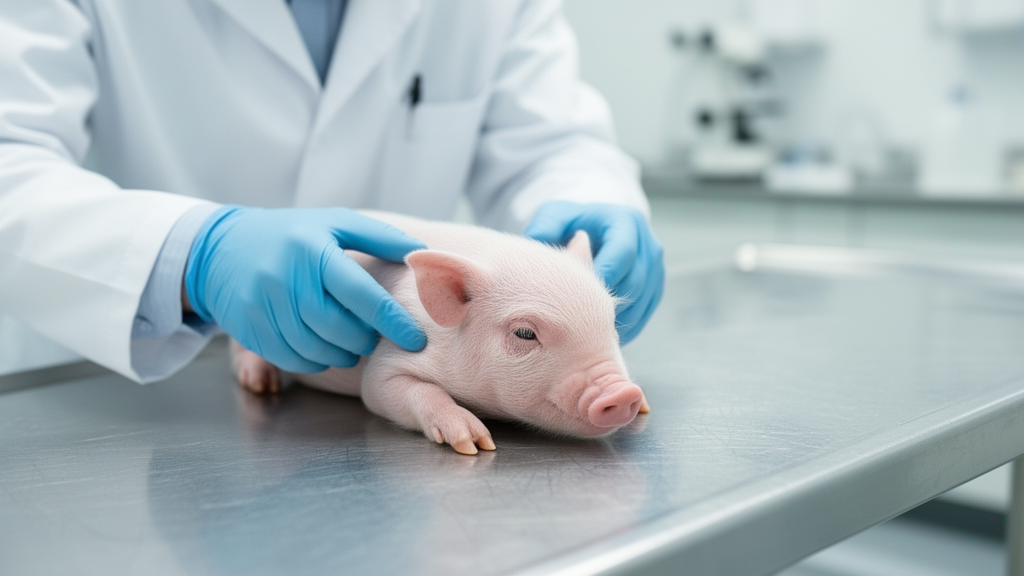
A detailed veterinary investigation conducted by researchers at the University of Copenhagen has demonstrated that macroscopic inspection of colonic content consistency and perianal faecal staining provides a practical and reproducible post-mortem indication of peri-mortem diarrhoea in young pigs. Published in the open-access journal Porcine Health Management, the study systematically evaluated whether anatomical observations made during necropsy could accurately reflect the diarrhoeic status of early-life piglets prior to death. Identifying whether an animal suffered from diarrhoea before dying is a frequent and critical challenge for veterinary pathologists and herd clinicians seeking to establish cause of death and understand disease dynamics in commercial swine production. Clinical signs of diarrhoea in live pigs are typically evaluated through pen-floor faecal pool inspections or direct rectal sampling, but these ante-mortem techniques depend heavily on the immediate availability of faecal material, which is often completely absent during routine post-mortem examinations. By contrasting specific luminal intestinal content characteristics across multiple gastrointestinal segments against standard reference metrics, the research team established that colonic fluid dynamics correlate strongly with physiological fluid loss while other intestinal segments offer negligible diagnostic value.
The study cohort comprised 145 crossbred piglets selected from a specific pathogen-free commercial indoor swine herd in Denmark that was documented free of major systemic infections including pleuropneumonia, swine dysentery, atrophic rhinitis, mange, lice, and porcine reproductive and respiratory syndrome. The monitored animals were three-way crosses produced from Landrace and Yorkshire sows inseminated with mixed semen from Duroc boars. Litters were monitored longitudinally from birth through ten weeks of age, with weaning occurring at twenty-six days of age before piglets were randomised into nursery accommodation. To capture developmental changes and varying disease manifestations across the early life period, pigs were assigned to age-stratified batches and euthanised at precise developmental milestones, specifically targeting twenty piglets at four days of age, twenty-five piglets at fourteen days, twenty-five piglets at twenty-five days, forty-five piglets at forty-six days, and thirty piglets at sixty-seven days. Antibiotic administrations on the farm were restricted solely to individual clinical indications without any routine metaphylactic batch treatments, providing an unmanipulated baseline to examine natural variations in gastrointestinal pathology and fluid accumulation.
To obtain reliable post-mortem specimens, sampling sessions were scheduled across eleven consecutive batches between July 2023 and March 2024, with each group transported gently in soft bedding for approximately one and a half hours from the commercial facility to the university necropsy laboratories. Immediately preceding euthanasia, clinical faecal scores were recorded using the four-point cotton-swab technique inserted into the rectum to capture adhering material. Following intramuscular anaesthesia with Zoletil and terminal intracardiac pentobarbital overdose, examiners recorded the presence or absence of external perianal faecal staining on the perineum and ventral tail surface through consensus agreement. During evisceration completed strictly within twenty minutes of death, five distinct gastrointestinal segments—the duodenum, jejunum, ileum, caecum, and colon—were isolated using haemostatic clamps to prevent fluid shifting, opened longitudinally, pinned onto boards, and systematically photographed. Luminal contents from each anatomical segment were categorised using standardised visual score sheets spanning consistency and colour, while descending colonic content was frozen for definitive gravimetric dry-matter determination via controlled microwave evaporation, and rectal swabs were analysed via microfluidic real-time polymerase chain reaction.
Quantitative assessment of observer agreement revealed striking differences in the reproducibility of physical consistency scoring compared to visual colour scoring across the gastrointestinal tract. For content consistency, the inter-rater reliability evaluated through Krippendorff’s alpha coefficient was remarkably robust, exceeding 0.80 across almost all examined intestinal segments and reaching an alpha value of 0.815 within the colon, with the caecum representing the only segment demonstrating moderate rather than high agreement. When raters were tasked specifically with determining the categorical presence or absence of diarrhoea based on colonic content appearance, unanimous consensus among all independent evaluators was achieved in ninety-two percent of all examined cases. In stark contrast, visual scoring of luminal content colour exhibited poor inter-rater reproducibility across all intestinal segments, yielding Krippendorff’s alpha values consistently below 0.64. While this colour agreement remained somewhat above chance expectation, the marked observer subjectivity indicates that visual colour grading cannot serve as an objective standalone metric in field post-mortem examinations without substantial risk of misclassification.
When diagnostic indicators were evaluated against gravimetric faecal dry-matter content in post-weaning piglets, colonic content consistency demonstrated high diagnostic sensitivity but limited specificity. Classifying colonic contents exhibiting mucoid, runny, or watery appearances as indicative of diarrhoea achieved a sensitivity of 86.7 percent and an overall diagnostic agreement of 69 percent when compared against dry-matter thresholds. However, colonic scoring yielded a comparatively low specificity of 55.3 percent, driven primarily by a consistent unidirectional discrepancy where visual colonic scoring assigned a score exactly one point higher than the corresponding dry-matter measurement. Samples classified as score two by dry-matter analysis were frequently categorised as score three during visual colonic inspection, resulting in an overestimation of diarrheic cases. The authors noted that this phenomenon aligns with normal digestive physiology, as the distal colon represents the terminal site of active water and electrolyte absorption, meaning luminal content situated further upstream macroscopically naturally contains higher fluid proportions than fully formed faecal matter.
External examination of perianal faecal staining emerged as another strongly correlated macroscopic indicator of severe gastrointestinal fluid discharge, though its diagnostic performance similarly declined when applied to subtle clinical variations. Visible perianal staining was recorded in thirty-six percent of the total cohort, representing 52 of 145 animals, but appeared in only five piglets prior to weaning. The statistical association between colonic consistency classifications and the presence or absence of perianal staining was highly significant (P = 0.0005). Remarkably, animals presenting with pelleted or hard paste colonic contents never exhibited perianal faecal staining, whereas one hundred percent of piglets with watery colonic contents displayed pronounced external staining. However, post-weaning diagnostic agreement between perianal staining and the rectal cotton-swab method fell to fifty-four percent. While visible perineal staining reliably differentiated severely diarrhoeic piglets from completely unaffected individuals, it proved incapable of discriminating between mild and intermediate consistency categories, limiting its utility for detecting early-stage or mild enteric pathology during routine disease surveillance.
In contrast to the valuable diagnostic insights provided by the large intestine, content consistency within the jejunum demonstrated complete absence of utility for detecting peri-mortem diarrhoea. Although small intestinal disease processes are frequently implicated in the clinical onset of early-life diarrhoea, statistical analysis using Kendall’s tau showed no meaningful correlation between jejunal luminal consistency and ante-mortem cotton-swab faecal scores in either suckling or post-weaning piglets. Even when severe watery diarrhoea was confirmed downstream, jejunal chyme fluidity failed to reflect rectal or colonic consistency. Consequently, veterinary pathologists evaluating piglet carcasses cannot rely on the viscosity, fluidity, or macroscopic appearance of small intestinal contents to infer whether an animal suffered from diarrhoea prior to death. Furthermore, the ileum was found to be entirely empty in a substantial proportion of animals across all age groups, severely restricting the practical feasibility of performing systematic morphological evaluations on luminal contents in that segment during standard necropsy workflows.
The study also revealed substantial shortcomings and bidirectional variations when assessing the cotton-swab scoring technique against laboratory-determined faecal dry-matter percentages. Although the cotton-swab method and dry-matter determinations identified nearly identical numbers of diarrhoeic animals in the post-weaning group—recording thirty-one and thirty cases respectively—the specific individual animals identified by the two methods failed to match in thirty-three percent of cases. When individual score discrepancies were analysed, differences between the cotton swab and dry-matter reference were frequently wide and bidirectional, with seven cases deviating by three full points on the four-point scale. Such profound discrepancies could stem from diurnal biological fluctuations in faecal moisture, intermittent shedding, or intrinsic scoring inaccuracies when minimal sample volumes adhere to the swab. Because the cotton-swab technique has never been formally validated in pre-weaning piglets and lacks published interobserver reliability data, these findings underline the danger of relying unquestioningly on swab scoring as an infallible clinical reference.
Molecular screening of rectal swabs using high-throughput microfluidic real-time polymerase chain reaction identified several statistically significant correlations between intestinal content colour and specific enteric pathogens, though these findings must be viewed through the lens of observer subjectivity. Dark coloration of the ileal, caecal, and colonic contents was linked to pathogen presence; in the colon, dark contents showed marked relative risk associations with Lawsonia intracellularis (RR = 22.3) and Brachyspira pilosicoli (RR = 18.4). In the caecum, dark colour was associated with B. pilosicoli (RR = 8.2), while ileal dark colour correlated with L. intracellularis (RR = 6.8). In the jejunum, detection of Clostridium perfringens alpha and beta2 toxin genes occurred almost exclusively in yellow contents, while in the colon, green coloration carried an elevated risk of clostridial toxins (RR = 5.7 to 12.0) and dark coloration exhibited a protective association (RR = 0.06 to 0.07). Nonetheless, because intestinal colour naturally transitions from pale milk-associated hues to darker post-weaning tones as diets change, these pathogen associations are heavily confounded by age and feeding transitions.
In their concluding synthesis, the authors emphasized the practical clinical implications and notable constraints of their findings for on-farm disease management and necropsy diagnosis. Because macroscopic colonic scoring and perianal staining both suffer from limited diagnostic specificity when judged against dry-matter standards, veterinary assessments relying solely on gross inspection risk inflating the apparent prevalence of diarrhoea through false-positive classifications. In commercial production, such diagnostic overestimation could trigger unnecessary antimicrobial interventions at the batch level, running counter to European regulatory goals aimed at minimizing therapeutic antibiotic usage following bans on medicinal zinc oxide and growth promoters. The authors cautioned that their study was conducted within a single commercial herd using qualitative rather than quantitative pathogen detection, and pre-necropsy transport handling may have contributed to acute stress-induced bowel evacuation. To overcome visual subjectivity, future research must incorporate multi-herd cohorts, quantitative pathogen load profiling, and automated digital image analysis or artificial intelligence tools to achieve standardized, objective post-mortem enteric diagnoses.
Subject of Research: Diagnostic accuracy of macroscopic post-mortem indicators and intestinal content morphology for detecting diarrhoea in young pigs
Article Title: Comparative evaluation of post-mortem methods for detection of diarrhoea in early-life piglets
Article References: Becker, C. B., Morsing, M. K., Laursen, J. R., Hansen, E. A. K., Nielsen, J. P., Fredholm, M., Goecke, N. B., Hansen, M. S., Nielsen, S. S., & Jensen, H. E. (2026). Comparative evaluation of post-mortem methods for detection of diarrhoea in early-life piglets. Porcine Health Management, 12(1), Article 42. https://doi.org/10.1186/s40813-026-00523-3
Image Credits: AI Generated
DOI: 10.1186/s40813-026-00523-3
Keywords: Porcine Health Management, piglets, diarrhoea, post-mortem examination, necropsy indicators, colonic content consistency, perianal faecal staining, cotton swab method, faecal dry matter, jejunal content, Krippendorff alpha, interobserver reliability, Lawsonia intracellularis, Brachyspira pilosicoli, Clostridium perfringens, real-time PCR, post-weaning diarrhoea, veterinary pathology
News Source: Ophelia Keating. (November 19, 2026). Post-Mortem Colonic Consistency Effectively Identifies Peri-Mortem Diarrhoea in Early-Life Piglets. Scienmag.