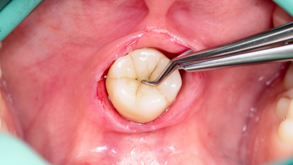
A surgical approach using a lingual mucoperiosteal flap achieved successful intraoral retrieval of iatrogenically displaced mandibular third molars and root fragments in all 18 consecutive patients evaluated in a retrospective clinical study. Dislodgement of a lower wisdom tooth or its root portion into the adjacent anatomical floor of the mouth represents an uncommon but clinically serious complication of third molar dental extractions. Prior published literature describing this surgical complication has remained largely confined to isolated case reports, leaving procedural management unstandardised across dental practices.
The retrospective evaluation was conducted at Art Dentist Dental Clinic, a private oral surgery clinic located in Hanoi, Vietnam, spanning clinical treatments and follow-up care carried out between January 2023 and December 2025. Consecutive patients were enrolled in the investigation if the displacement of mandibular third molars or root fragments into the sublingual or submandibular spaces was verified through clinical examination and cone-beam computed tomography imaging. All participants underwent retrieval using the same standardised lingual mucoperiosteal flap technique on the day of presentation and completed a minimum follow-up duration of six months.
Within the evaluated cohort of 18 patients, the mean age was recorded as 26.72 ± 5.88 years, with male patients comprising a slight majority of 55.56 percent. The overwhelming majority of the dislodged dental structures were roots rather than complete teeth, accounting for 94.44 percent of cases, with a measured median fragment size of 4.00 millimetres. The documented clinical histories revealed that 88.89 percent of the accidental displacements originally occurred during extraction procedures performed by general dental practitioners prior to referral.
Diagnostic assessment evaluated the presence of the radiographic Y-sign, an anatomical feature identified on panoramic radiographs whose diagnostic utility in predicting or identifying displacement risk has historically remained unclear. Among the subgroup of seven patients who had available pre-extraction panoramic radiographs, the Y-sign was observed in every individual, representing a 100 percent detection rate within that evaluated subset. In addition to examining radiographic characteristics, the investigation noted the frequency of preoperative anxiety among presenting patients awaiting surgical retrieval.
Surgical intervention yielded complete procedural success across the entire study sample, with successful retrieval documented in all 18 patients, corresponding to a success rate of 100 percent with a 95 percent confidence interval spanning from 81.47 to 100 percent. The operations were conducted on the day of initial presentation, even for individuals presenting with small anatomical fragments. The procedures demonstrated a median operating time of 22.50 minutes from initiation to completion of the retrieval process.
Postoperative neurological assessments focused on evaluating potential sensory complications following manipulation near lingual structures. Non-blinded pin-prick testing detected no newly acquired neurosensory deficits following the operation, yielding a new deficit rate of zero percent with a 95 percent confidence interval between 0.00 and 18.53 percent. In two individuals who presented with pre-existing neurosensory deficits sustained prior to the retrieval intervention, the nerve impairments achieved complete clinical resolution within five months of follow-up.
The researchers underscored several methodological boundaries inherent to the study design, noting that the data originated from a single-centre retrospective cohort lacking a parallel comparison group. Because of these constraints, the investigators stated that the reported outcomes and the radiographic Y-sign findings must be interpreted as exploratory and hypothesis-generating observations rather than definitive practice standards. The authors concluded that larger prospective clinical studies are necessary before the approach can be broadly adopted across routine surgical settings.
Subject of Research: Intraoral surgical retrieval of displaced mandibular third molars using a lingual mucoperiosteal flap
Article Title: Intraoral retrieval of iatrogenically displaced mandibular third molars from the sublingual and submandibular spaces using a lingual mucoperiosteal flap: a retrospective study of 18 patients
Article References: Nguyen, X. T., Pham, T. H., Hoang, N. L., Huynh, N. D., & Nguyen, T. T. (2026). Intraoral retrieval of iatrogenically displaced mandibular third molars from the sublingual and submandibular spaces using a lingual mucoperiosteal flap: a retrospective study of 18 patients. BMC Oral Health. https://doi.org/10.1186/s12903-026-10451-x
Image Credits: AI Generated
DOI: 10.1186/s12903-026-10451-x
Keywords: mandibular third molar, iatrogenic displacement, lingual mucoperiosteal flap, oral surgery, tooth extraction complication, root fragment retrieval, sublingual space, submandibular space, cone-beam computed tomography, Y-sign, retrospective study, neurosensory deficit, pin-prick testing, dental surgery, operating time, Hanoi Vietnam
News Source: Ophelia Keating. (November 18, 2026). Lingual Flap Approach Successfully Retrieved Displaced Third Molars in Retrospective Study. Scienmag.