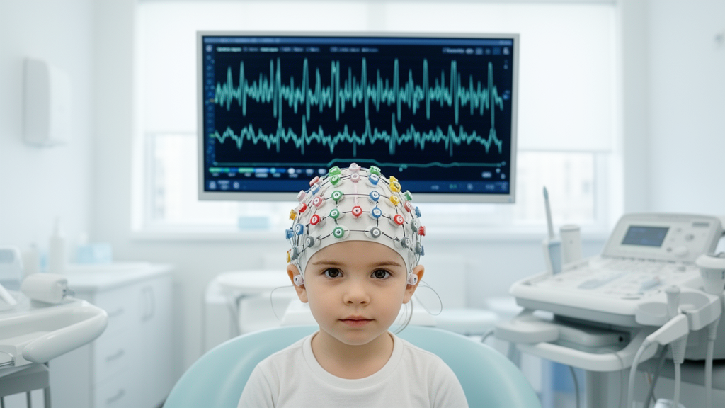
In an extensive single-center investigation evaluating cortical electrical activity, researchers identified clinically abnormal electroencephalographic patterns in more than half of assessed children diagnosed with autism spectrum disorder, uncovering distinct clinical profiles that correlate with specific neuroanatomical brain regions. The retrospective observational study, conducted at Peking University Sixth Hospital and analyzing 343 pediatric patients assessed between January 2022 and May 2024, demonstrated that overall electroencephalographic abnormalities were present in 53.4 percent of participants. Multivariable modeling established that a documented history of developmental regression, an age between 4 and 5.9 years, and premature birth were independently associated with higher odds of exhibiting abnormal electroencephalographic activity. Crucially, exploratory subgroup analyses revealed that these risk profiles varied markedly across anatomical locations, indicating that distinct clinical characteristics map onto electrophysiological disturbances in specific cortical lobes rather than displaying uniform distribution throughout the cerebrum.
The study addresses a longstanding challenge in developmental neuropsychiatry regarding the co-occurrence of electrophysiological disturbances and neurodevelopmental conditions. Autism spectrum disorder is characterized by persistent challenges in social communication alongside restricted and repetitive patterns of behavior, interests, or activities. A documented comorbidity is epilepsy, with literature estimating prevalence rates between 10 percent and 46 percent among autistic individuals, substantially exceeding general population rates. Visual inspection of electroencephalography by qualified experts serves as the clinical standard for identifying epileptiform activity, yet clinicians increasingly recognize that many children with autism display abnormal electroencephalographic features without experiencing clinical seizures. Emerging literature suggests these subclinical discharges may relate to cognitive deficits, language impairments, or behavioral manifestations such as irritability and aggression. However, previous studies assessing risk factors have yielded inconsistent findings, rarely evaluating whether identified clinical variables differentially influence distinct neuroanatomical locations where abnormal electrical activity originates.
To rigorously investigate whether clinical predictors exert differential effects across neuroanatomical locations, the research team retrospectively evaluated pediatric patients diagnosed with autism spectrum disorder at Peking University Sixth Hospital. Diagnoses were systematically confirmed by experienced child psychiatrists following the Diagnostic and Statistical Manual of Mental Disorders, Fifth Edition diagnostic criteria. Detailed prenatal, perinatal, and postnatal medical records were extracted from hospital charts to characterize each child across demographic, family, and developmental domains. To isolate electrophysiological patterns from confounding neurological events, the investigators excluded individuals presenting with acute neurological conditions such as traumatic brain injury, children with established prior diagnoses of overt epilepsy, and individuals receiving pharmacotherapies known to alter brainwave signals, such as antiepileptic medications. Furthermore, recruitment was not restricted to children undergoing examination for clinically suspected seizures; caregivers of all age-eligible autistic children were offered voluntary participation as part of comprehensive neurodevelopmental evaluations.
From an initial pool of 823 children with available electroencephalographic recordings, clinical exclusions eliminated 62 individuals due to acute neurological disorders or medication use that directly affects neurophysiological signals. An additional 337 children were excluded because of incomplete child or family background documentation, and 81 were removed due to incomplete developmental characteristics, yielding a final analytical cohort of 343 children. Methodological comparisons between the 343 included participants and the 418 children excluded for incomplete data revealed an age difference, with the analyzed cohort being younger, presenting a mean age of 5.13 years with a standard deviation of 2.61 years compared to 6.69 years with a standard deviation of 4.00 years among excluded peers. The proportion of male children remained similar across groups, comprising 79.6 percent of the analyzed cohort and 81.3 percent of the excluded cohort. Overall, the final sample consisted predominantly of boys and presented an average age of 5.1 years.
The technical protocol employed non-sedated, long-term video-electroencephalography monitoring utilizing a 128-channel NicoletOne recording system with scalp electrodes configured according to the international 10–20 placement system. Recordings utilized an average reference montage with a sampling frequency of 512 hertz and bandpass filtering configured between a low-cut threshold of 0.5 hertz and a high-cut threshold of 70 hertz. Every included child underwent continuous recording lasting at least four hours without sedation, avoiding pharmaceutical alterations of background rhythms while substantially increasing the probability of capturing natural sleep, which is recognized to enhance the detection of epileptiform discharges. Successful natural sleep recordings were captured in 312 of the 343 participants, representing 91.0 percent of the cohort. Dedicated electrophysiology technicians performed recordings upon hospital presentation, and two board-certified neurophysiologists evaluated recordings blinded to all clinical and developmental information, ensuring objective waveform classification according to standard institutional clinical reporting frameworks.
Electroencephalographic recordings were designated as clinically abnormal whenever at least one pathological waveform abnormality was documented during routine clinical interpretation. The primary outcome captured both epileptiform discharges, including spikes and sharp waves, and non-epileptiform pathological abnormalities, such as background slowing where theta or delta frequencies exceeded 50 percent of background activity. Atypical spindle-like activity outside non-rapid eye movement stage 2 sleep was coded as spindle-wave abnormalities, whereas physiological sleep spindles were excluded. The primary outcome was not restricted to epileptiform events or epilepsy diagnoses. Topographical categories encompassed frontal, temporal, parietal, and occipital lobes, alongside a multi-region category defined as abnormalities simultaneously involving two or more anatomical regions. These regional indicators were not mutually exclusive, allowing a child exhibiting focal discharges across multiple lobes to contribute data to each respective region-specific model. Generalized involvement, documented in 111 children representing 32.4 percent of the cohort, was analyzed as an independent topographic category.
Statistical evaluation utilized multivariable logistic regression modeling to assess independent predictors of overall and region-specific electroencephalographic abnormalities. To control for confounding influences across multiple life stages, every adjusted regression model incorporated 12 prespecified covariates spanning perinatal, family, and developmental domains, estimating 13 distinct regression parameters excluding the model intercept. Perinatal covariates included caesarean delivery, birth asphyxia, and premature birth defined as gestational age under 37 weeks. Family characteristics comprised family histories of mental illness across first-degree and second-degree relatives, advanced maternal age at delivery defined as 35 years or older, documented threatened abortion, and maternal gestational diseases such as gestational diabetes or hypertension. Developmental variables captured documented co-occurring intellectual disability, attention-deficit/hyperactivity disorder, and developmental regression. Regression was operationally defined as documented caregiver reports during clinical interviews showing a clear loss of previously acquired language or social-communication skills following normal developmental progress prior to age six.
In the primary multivariable regression model examining overall electroencephalographic status, abnormal recordings were identified in 183 children, establishing an abnormal prevalence of 53.4 percent compared to 160 normal recordings. Documented developmental regression demonstrated the strongest statistical association with overall abnormalities, yielding an adjusted odds ratio of 6.53 with a 95 percent confidence interval spanning 1.79 to 23.72. Children in the preschool developmental bracket of 4 to 5.9 years exhibited significantly elevated odds of abnormal recordings compared to the youngest reference group aged 2 to 3.9 years, producing an odds ratio of 2.46 with a 95 percent confidence interval of 1.38 to 4.37. Preterm birth also showed a significant independent association, displaying an odds ratio of 2.54 with a 95 percent confidence interval of 1.00 to 6.43. Attention-deficit/hyperactivity disorder exhibited a positive trend with an odds ratio of 2.28 and a confidence interval of 0.93 to 5.60, whereas sex, caesarean delivery, birth asphyxia, family mental illness history, advanced maternal age, maternal pregnancy diseases, threatened abortion, and intellectual disability demonstrated no statistically significant associations.
Exploratory region-specific analyses revealed distinct anatomical distributions and localized predictor associations across cerebral lobes. Among the analyzed children, frontal abnormalities were documented in 55 participants representing 16.0 percent, temporal abnormalities occurred in 45 children representing 13.1 percent, parietal findings appeared in 37 children representing 10.8 percent, occipital abnormalities were identified in 13 children representing 3.8 percent, and multi-region abnormalities were detected in 56 children representing 16.3 percent. Multivariable modeling of frontal lobe abnormalities demonstrated significant independent associations with attention-deficit/hyperactivity disorder, yielding an odds ratio of 3.75 with a 95 percent confidence interval of 1.41 to 9.99, developmental regression with an odds ratio of 3.89 and a confidence interval of 1.43 to 10.56, and premature birth with an odds ratio of 2.71 and a confidence interval of 1.04 to 7.08. Conversely, temporal lobe abnormalities were uniquely associated with developmental regression, producing an odds ratio of 3.51 with a confidence interval of 1.27 to 9.75, while no other assessed variable showed a significant relationship. Multi-region involvement was significantly associated solely with premature birth, showing an odds ratio of 2.77 and a confidence interval of 1.08 to 7.11.
These topographical differentiations offer biologically plausible hypotheses regarding distinct neural networks implicated in autism spectrum disorder. The temporal lobe is essential for language acquisition, auditory processing, and social cognition; when abnormal electrical discharges disrupt temporal networks, the localization aligns closely with the loss of acquired language and social communication characteristic of developmental regression. In contrast, the frontal lobe governs executive functioning, impulse regulation, and sustained attentional control. The co-occurrence of attention-deficit/hyperactivity disorder, prematurity, and frontal electroencephalographic abnormalities aligns with known vulnerabilities in frontostriatal and frontoparietal cognitive circuits following altered early cortical development. Furthermore, the peak prevalence of abnormalities observed in preschool children aged 4 to 5.9 years may delineate a sensitive developmental window marked by intensive synaptic remodeling and neural circuit reorganization, during which aberrant synchronization becomes most detectable macroscopically before potential maturation-related normalization in later childhood stages.
Notwithstanding these robust statistical observations, the authors emphasize essential methodological limitations that restrict immediate clinical translation. The study relied on a retrospective cross-sectional design, precluding causal inferences and leaving open the possibility that observed associations reflect non-causal neurodevelopmental correlates. Several regional outcomes, particularly occipital lobe abnormalities with only 13 recorded events, contained limited event counts relative to the 13 parameters evaluated per model, increasing susceptibility to overfitting and yielding wide confidence intervals that require treating regional findings as hypothesis-generating. Selection bias could not be entirely excluded because excluded children were older than the analyzed cohort. Furthermore, developmental regression was documented retrospectively from parental reports in medical records, yielding a cohort prevalence of 6.1 percent that likely underrepresented milder regressions compared to literature benchmarks around 30 percent. Routine genetic testing was unavailable, leaving potential residual confounding from unmeasured syndromic conditions. Consequently, the investigators caution that these electrophysiological associations do not demonstrate that routine electroencephalographic screening improves clinical management or individual patient outcomes, highlighting instead the necessity for prospective, multicenter longitudinal studies to track neurodevelopmental trajectories over time.
Subject of Research: Topography-specific risk factors for electroencephalographic abnormalities in children with autism spectrum disorder
Article Title: Topography-specific risk factors for EEG abnormalities in children with autism spectrum disorder: insights from long-term video-EEG monitoring
Article References: Zhao, Y., Jia, M., Bai, Q., Wang, R., Wang, H., & Jia, M. (2026). Topography-specific risk factors for EEG abnormalities in children with autism spectrum disorder: insights from long-term video-EEG monitoring. BMC Psychiatry. https://doi.org/10.1186/s12888-026-08861-w
Image Credits: AI Generated
DOI: 10.1186/s12888-026-08861-w
Keywords: autism spectrum disorder, electroencephalography, video-EEG monitoring, developmental regression, prematurity, ADHD comorbidity, frontal lobe abnormalities, temporal lobe abnormalities, pediatric neurology, neurodevelopmental disorders, epileptiform discharges, epilepsy comorbidity, cross-sectional study, cortical function, topographical distribution, odds ratio, sleep EEG, multivariable logistic regression
News Source: Glenn Wilkins. (November 18, 2026). Study Links Specific Clinical Profiles to Distinct EEG Abnormalities in Autistic Children. Scienmag.