In an exploratory investigation evaluating the genetic architecture of human facial variation, researchers have identified significant associations between common single nucleotide polymorphisms in the fibroblast growth factor receptor 2 gene, known as FGFR2, and distinct craniofacial dimensions in children. The cross-sectional investigation, conducted using digital photogrammetric records and genomic DNA collected from saliva, revealed that specific functional polymorphic alleles correlate with measurable variations in eye spacing, midface balance, lower lip vermilion proportions, and the profile contour of the lower jaw. Conducted across a pediatric cohort evaluated prior to the initiation of orthodontic therapy, the research provides preliminary empirical evidence that common genetic variants within signaling pathways previously linked to severe craniofacial malformations also contribute to normal phenotypic differences across healthy individuals.
The human face serves as a primary determinant of interpersonal recognition, social perception, and morphological identity, emerging through an intricate morphogenetic program regulated by numerous environmental and genetic factors. Historically, the profound role of heredity in establishing facial shape and dimension has been supported by observations of familial resemblances and high concordance rates in monozygotic twin pairs. Over the past two decades, genomic research has increasingly focused on dissecting the specific loci that govern subtle morphological traits across the skull and soft tissue contours. Particular attention has centered on fibroblast growth factor receptor 2, a key signaling protein essential for normal embryonic craniofacial morphogenesis and suture homeostasis. Severe gain-of-function and loss-of-function mutations within FGFR2 are known causative drivers of rare syndromic craniosynostoses, such as Crouzon, Apert, and Pfeiffer syndromes, in which premature cranial suture fusion produces characteristic dysmorphic alterations, including prominent midface hypoplasia and orbital proptosis. Because polymorphic variations occurring at population frequencies of one percent or higher can modulate gene expression levels or alter functional protein structures, the investigators hypothesized that common FGFR2 polymorphisms might also underpin non-pathological variations in the facial structures of healthy human populations.
To investigate this genotype-phenotype relationship, the research team designed and conducted an exploratory association study structured in accordance with the Strengthening the Reporting of Genetic Association study, or STREGA, checklist. The project was embedded within an orthodontic clinic setting in Regensburg, Germany, where patients were recruited between January 2020 and June 2021. Restricting enrollment to prospective orthodontic candidates before the placement of corrective appliances ensured that soft tissue morphology remained uninfluenced by mechanical tooth movement or subsequent orthodontic repositioning of the dental arches. All participating children and their families shared similar European ancestral backgrounds, with ancestral continuity verified across parents and grandparents through self-declared lineage. This rigorous geographic and demographic matching was instituted to minimize potential confounding effects arising from population stratification, which can introduce spurious associations in candidate gene investigations when diverse sub-populations exhibit differing baseline allele frequencies and morphological norms.
Statistical sample size considerations were established prior to subject enrollment, using statistical power modeling designed around the lowest expected minor genotype frequency among the candidate FGFR2 polymorphisms, set at 0.15. Anticipating a mean facial measurement difference of 0.09 with an estimated standard deviation of 0.1 across genotype groups based on previously reported facial morphology data in distinct populations, the calculation established that a minimum sample of 73 individuals was necessary to achieve 80 percent statistical power at a two-sided alpha threshold of 0.05. Stringent clinical exclusion criteria were implemented to isolate non-syndromic variation: children diagnosed with systemic syndromes, individuals with a clinical history of significant skull trauma, patients presenting with cleft lip or palate, and individuals with congenital tooth agenesis outside third molars were systematically excluded from the investigation. Additionally, potential subjects with incomplete documentation, substandard photographic clarity, or inadequate saliva specimens were eliminated from the analytical pipeline.
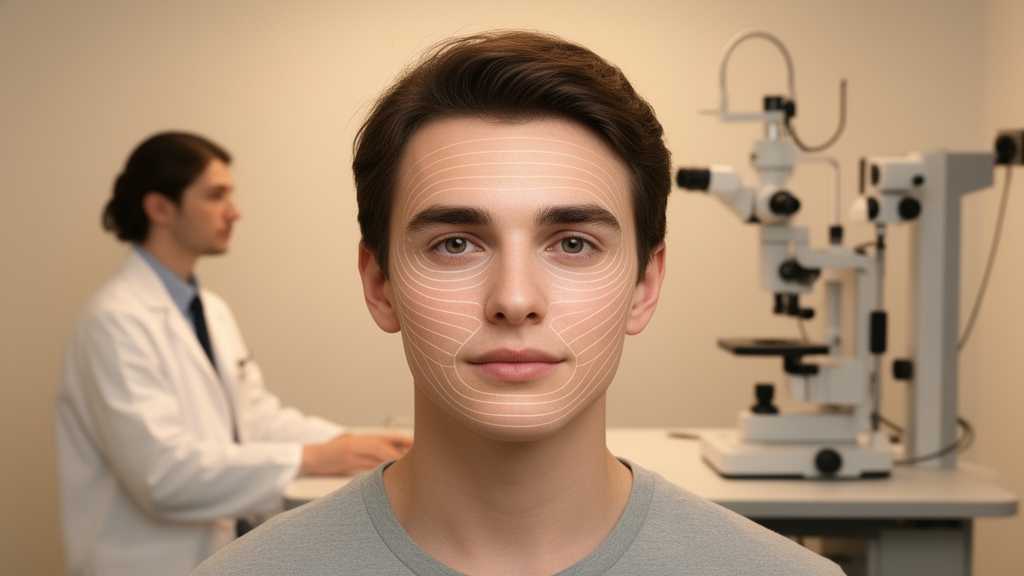
Phenotypic characterization was conducted through standardized two-dimensional digital photogrammetry using pre-existing digital frontal and lateral facial profile photographs taken under controlled clinical settings. To ensure reliable anatomical tracking, the research team identified 29 soft tissue landmarks distributed across frontal and profile views, selected specifically for their established visibility and high anatomical reproducibility in published literature. Frontal images were aligned using the horizontal bipupillary line connecting the right and left pupils as a standard reference axis, whereas lateral profile images were standardized along the Frankfurter horizontal line, drawn from the inferior margin of the orbit to the porion at the superior edge of the external auditory meatus. Because clinical photographic images were captured without a physical millimeter calibration scale, measurements were converted into proportional ratios and angular relations rather than absolute linear units, calculating pixel distances within the ImageJ software platform. Intra-examiner reliability for landmark placement and coordinate quantification was verified by having a single calibrated evaluator re-measure a subset of ten frontal and ten profile photographs after a two-week interval, yielding intraclass correlation coefficients ranging from 0.93 to 0.99.
From these digital anatomical coordinates, the evaluators derived 22 discrete linear proportions and angular measurements for each enrolled child. Frontal photographic relationships assessed transverse midfacial dimensions, including the ratio of the inner intercanthal distance to the outer intercanthal distance, the ratio of the inner intercanthal distance to the pupillary midface span, the proportion of nasal base width relative to overall alar width, and the proportion of lip height relative to transverse mouth width. Lateral profile relationships evaluated the relative vertical partitioning of the superior, middle, and inferior facial thirds along the vertical cranial plane, alongside detailed anterior lip projections and soft tissue chin metrics, such as the relative vermilion exposure of the upper and lower lips and the vertical height of the chin relative to the lower facial third. Biological samples were acquired through non-invasive saliva collection, from which genomic DNA was extracted from exfoliated oral epithelial cells. Following yield and purity quantification via spectrophotometry on a NanoDrop 1000 instrument, real-time polymerase chain reaction genotyping was conducted blindly using customized TaqMan assays on a StepOnePlus platform across seven candidate FGFR2 single nucleotide polymorphisms exhibiting minor allele frequencies exceeding 20 percent: rs2162540, rs2981578, rs10736303, rs1078806, rs11200014, rs1219648, and rs4752566.
The analytical cohort comprised 102 healthy children, consisting of 60 boys aged 9 to 14 years and 42 girls aged 9 to 13 years. The distinct age windows for male and female participants were deliberately chosen to reflect the documented developmental reality that female pediatric populations undergo pubertal growth spurts approximately one calendar year earlier than their male counterparts. Statistical evaluations of facial dimensional ratios and angular features demonstrated no statistically significant differences between genders across the cohort, allowing for unified genetic association modeling. Furthermore, genotyping call rates exhibited high technical reproducibility, ranging between 86.3 percent and 96.1 percent across the seven investigated loci. Goodness-of-fit testing confirmed that all observed genotype distributions conformed closely to expected Hardy-Weinberg equilibrium thresholds, with non-significant chi-square values indicating an absence of substantial sampling distortion, systemic genotyping error, or strong non-random mating patterns within the recruited European cohort.
Continuous phenotypic variables were evaluated across categorical genotype classifications using one-way analysis of variance and complementary independent-samples t-tests, adopting a standard significance criterion of alpha equal to 0.05 within the SPSS software environment. The statistical analysis revealed that the rs10736303 polymorphism in FGFR2 was significantly associated with relative transverse proportions across the eye and midfacial region. Specifically, the ratio comparing inner intercanthal distance to outer intercanthal distance demonstrated a statistically significant difference across genotypes (p = 0.013), and the ratio between inner intercanthal distance and pupillary midface distance showed a concordant significant association (p = 0.026). In both instances, individuals possessing the homozygous AA genotype exhibited larger relative midface and interocular proportions compared with heterozygous carriers possessing the AG genotype, suggesting that the presence of the A allele correlates with an expanded relative intercanthal space relative to surrounding orbitotemporal structures.
Lateral photographic profiling revealed additional phenotypic associations linked to FGFR2 genetic variation, particularly regarding the lower facial profile. The rs2162540 polymorphism demonstrated a statistically significant association with the relative vermilion height of the lower lip in relation to the inferior facial third (p = 0.049), with homozygous CC genotype carriers presenting a significantly reduced vermilion proportion compared to heterozygous CT individuals. Furthermore, lateral angular analysis highlighted a significant correlation between genetic polymorphisms and the mentolabial angle, defined by the soft tissue contour connecting the lower lip vermilion border, the supramentale point of deepest labiomental concavity, and the prominence of the soft tissue pogonion. Specifically, homozygous CC carriers of the rs2162540 variant exhibited a significantly larger mentolabial angle relative to CT individuals (p = 0.044). In parallel, the rs11200014 polymorphism demonstrated an independent association with the mentolabial angle (p = 0.024), where children homozygous for the AA genotype displayed significantly wider, more obtuse angles than heterozygous AG or homozygous GG carriers.
The biological convergence of these findings aligns with prior orthodontic literature demonstrating that FGFR2 polymorphisms correlate with skeletal Class II and Class III malocclusions, sagittal discrepancies that reflect underlying developmental mismatches between the maxilla and mandible, including mandibular retrognathism. Because variations in the mentolabial angle and relative lower lip posture represent recognized soft tissue manifestations of sagittal jaw positioning, the researchers reasoned that the observed phenotypic shifts may reflect FGFR2-mediated modulation of lower jaw growth, direct influences on soft tissue labial morphogenesis, or a combination of both developmental processes. The team highlighted several methodological limitations inherent to the study design, including the reliance on two-dimensional photographic ratios instead of three-dimensional stereophotogrammetry or millimeter-based direct anthropometry, the absence of hand-wrist or cervical vertebral radiographic imaging to verify peak growth timing, and the potential recruitment bias associated with sampling pre-orthodontic patients rather than an unselected general community cohort. Moreover, because 22 individual morphological traits were examined, the authors maintained an exploratory designation, deliberately foregoing conservative multiple testing corrections that would diminish statistical power and increase the probability of Type II false-negative errors. The findings establish a foundational baseline indicating that common functional variants within the FGFR2 pathway modulate normal pediatric facial contours, providing a rationale for future large-scale, three-dimensional population studies.
Subject of Research: Association between FGFR2 genetic polymorphisms and facial morphology in children
Article Title: Exploring the association between genetic polymorphisms in FGFR2 and children’s facial morphology
Article References: Rinnert, C., Beisel-Memmert, S., Proff, P., de Araujo, C. M., Baratto, M. J. P., Paiva Perin, C., Corá, J., Kirschneck, C., & Küchler, E. C. (2026). Exploring the association between genetic polymorphisms in FGFR2 and children’s facial morphology. Head & Face Medicine, 22(1), Article 67. https://doi.org/10.1186/s13005-026-00649-3
Image Credits: AI Generated
DOI: 10.1186/s13005-026-00649-3
Keywords: FGFR2, facial morphology, genetic polymorphisms, craniofacial development, rs10736303, rs2162540, rs11200014, mentolabial angle, intercanthal distance, vermilion height, orthodontic records, photogrammetry, ImageJ, Hardy-Weinberg equilibrium, craniosynostosis, European ancestry, skeletal malocclusion
News Source: Juliet Wilcox. (November 18, 2026). Common FGFR2 Gene Variants Linked to Distinct Facial Dimensions in Exploratory Study. Scienmag.