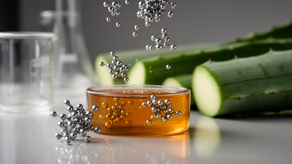
Researchers have synthesized stable, crystalline silver nanoparticles using an aqueous leaf extract of the century plant, Agave americana, acting simultaneously as a reducing and capping agent without requiring auxiliary chemical additives. In laboratory evaluations detailed in Health Nanotechnology, the resulting biogenic silver nanoparticles demonstrated broad-spectrum antibacterial efficacy against opportunistic Gram-negative and Gram-positive pathogens, measurable antioxidant radical-scavenging activity, and concentration-dependent cytotoxicity against cultured human SiHa cervical carcinoma cells. Physicochemical characterization confirmed the formation of predominantly spherical metallic nanoparticles stabilized by an organic biogenic corona composed of plant-derived biomacromolecules.
Conventional production pathways for nanoscale silver typically rely on chemical reduction reagents such as sodium borohydride or sodium citrate, or utilize physical techniques including laser ablation and arc discharge. Although effective at yielding uniform nanomaterials, these traditional methodologies frequently entail substantial energy consumption, generate hazardous chemical by-products, employ toxic synthetic precursors, and leave surface residues that compromise biological compatibility. To overcome these constraints, phyto-fabrication harnesses the intrinsic metabolic chemistry of plant tissues to reduce ionic silver to neutral elemental silver nuclei under mild laboratory conditions, providing an environmentally benign alternative that aligns with circular economy frameworks by utilizing renewable plant biomass.
The experimental protocol utilized fresh Agave americana foliage gathered from the campus of Babasaheb Bhimrao Ambedkar University in Lucknow, India. To extract active secondary metabolites, cleaned and dried leaf fragments were immersed in double-distilled water at a ratio of 4.5 grams per 100 milliliters and heated at 90 degrees Celsius for 20 minutes. Subsequent triple filtration yielded an optically clear brown aqueous filtrate enriched with phytochemicals, including steroidal saponins, phenolic acids, flavonoids, and complex polysaccharides. For the reduction stage, 4.5 milliliters of this freshly prepared extract was introduced into 200 milliliters of a 10 millimolar silver nitrate working solution maintained at 25 degrees Celsius under continuous orbital agitation at 150 revolutions per minute.
Following an initial 25-minute induction period, the mixture was transferred to 55 degrees Celsius and agitated at 500 revolutions per minute for 20 minutes, during which a characteristic brown-yellow hue developed in the liquid medium. The solution was held at this temperature for an additional 20 minutes to drive the reaction to completion before cooling to ambient room temperature. This colorimetric transition provides real-time optical verification of localized surface plasmon resonance, where conduction-band electrons confined within newly generated metallic clusters oscillate in resonance with incident light waves. Subsequent ultraviolet-visible absorption spectrophotometry confirmed a distinct absorption peak centered at 434 nanometers with a full width at half maximum of approximately 90 nanometers, and Tauc plot analysis indicated an effective optical bandgap of 2.32 electron volts.
Extensive structural analysis established the morphological and crystallographic features of the biosynthesized particles. Field-emission scanning electron microscopy of 760 individual nanoparticles revealed predominantly quasi-spherical profiles exhibiting smooth surface topographies, yielding an average physical diameter of 57 plus or minus 32.7 nanometers across a broader distribution extending from 20 to 800 nanometers. Powder X-ray diffraction recorded prominent reflection peaks at 2-theta values of 38.35, 44.01, 64.72, and 77.65 degrees, successfully indexing to the (111), (200), (220), and (311) lattice planes of face-centered cubic elemental silver cataloged in standard reference databases. Calculation via the Scherrer formulation established an average crystallite domain size of 19 nanometers, while the computed lattice parameter of 4.068 angstroms demonstrated close agreement with standard bulk silver parameters.
Colloidal behavior and surface functionalization were examined through dynamic light scattering, electrophoretic light scattering, and Fourier transform infrared spectroscopy. In aqueous dispersion, dynamic light scattering recorded an average hydrodynamic diameter of 68 nanometers alongside a polydispersity index of 0.21, demonstrating acceptable colloidal uniformity. Electrophoretic measurements established a zeta potential of minus 32.4 millivolts, a net negative surface charge governed by ionized carboxylate and hydroxyl functional groups residing within the biogenic capping layer. DLVO theoretical modeling indicated that this electrostatic surface potential creates an energy barrier of approximately 25 thermal energy units, providing sufficient electrosteric repulsion to suppress particle agglomeration during prolonged liquid storage.
Infrared spectral measurements substantiated the direct coordination of botanical components to the metallic silver core. Comparative analysis between the neat plant extract and the synthesized nanoparticles identified diagnostic vibrational shifts, most notably a displacement of the amide I band from approximately 1650 reciprocal centimeters in the raw extract down to 1631.96 reciprocal centimeters in the capped nanomaterial. Additional absorption bands appeared at 1033.95 reciprocal centimeters, 1292.06 reciprocal centimeters, and 800 reciprocal centimeters, matching carbon-oxygen stretching, amide III vibrations, and aromatic carbon-hydrogen deformations. These spectral signatures demonstrate that proteinaceous amides and polysaccharide hydroxyl groups derived from the leaf parenchyma assemble around the nascent silver core, forming a protective biogenic corona that prevents Ostwald ripening.
Biological screening revealed potent antimicrobial properties during agar disk diffusion testing against standard microbial strains, specifically Escherichia coli, Pseudomonas aeruginosa, and Staphylococcus aureus. Exposure to nanoparticle suspensions at 100 micrograms per milliliter produced distinct clear zones of growth inhibition measuring 18.0 plus or minus 0.2 millimeters for Escherichia coli, 16.7 plus or minus 0.1 millimeters for Staphylococcus aureus, and 14.5 plus or minus 0.2 millimeters for Pseudomonas aeruginosa. In broth dilution testing, the corresponding minimum inhibitory concentrations were 3.0 micrograms per milliliter for Escherichia coli, 5.0 micrograms per milliliter for Staphylococcus aureus, and 6.0 micrograms per milliliter for Pseudomonas aeruginosa, with the nanoparticles exhibiting inhibitory potency exceeding that of a 10-microgram commercial gentamicin control against Pseudomonas aeruginosa under identical test parameters.
In vitro antioxidant testing using the 2,2-diphenyl-1-picrylhydrazyl radical bleaching assay confirmed concentration-dependent free-radical quenching capability. The biogenic nanoparticles achieved 84.2 percent radical scavenging at a concentration of 100 micrograms per milliliter, yielding an overall half-maximal inhibitory concentration of 48 micrograms per milliliter, compared to 98.4 percent inhibition and an inhibitory concentration of 32 micrograms per milliliter recorded for standard ascorbic acid. The extensive surface area of the nanostructures, estimated at roughly 12.7 square meters per gram, concentrates functional plant phenolics and allows the metallic silver surface to facilitate electron-transfer processes, neutralizing stable organic free radicals in solution.
Cytotoxicity assessments performed via the colorimetric MTT assay across cultured SiHa human cervical carcinoma cells revealed dose-dependent inhibition of mitochondrial metabolic activity following 24 hours of exposure. The phyto-fabricated nanoparticles achieved a half-maximal inhibitory concentration of 15 micrograms per milliliter against the malignant cell line, displaying markedly higher cytotoxic potency than the unformulated crude Agave americana leaf extract, which yielded an inhibitory concentration of 30 micrograms per milliliter under the same assay conditions. The enhanced potency observed in the nanoformulation reflects increased cellular interactions and localized delivery of bioactive silver species facilitated by nanoscale physical dimensions.
Thermal analysis revealed important composition characteristics alongside key analytical constraints. Differential scanning calorimetry registered a prominent endothermic transition at 103.88 degrees Celsius, corresponding to the loss of surface-adsorbed moisture, while thermogravimetric analysis initiated major mass reduction near 112 degrees Celsius as organic stabilizers underwent progressive thermal breakdown. However, total mass loss over the 25 to 800 degrees Celsius thermal scanning profile reached approximately 99.1 percent. This substantial mass reduction indicates that the tested lyophilized sample possessed a high proportion of organic extract matrix rather than pure metallic silver, meaning these specific thermal curves reflect the decomposition of botanical constituents rather than the intrinsic core thermal stability of isolated silver nanoparticles.
The study authors emphasize that these experimental findings represent preliminary in vitro evaluations that warrant careful interpretation. While the biogenic silver nanoparticles demonstrated robust antimicrobial, antioxidant, and cytotoxic activities, the precise biological modes of action—such as intracellular reactive oxygen species generation, membrane disruption, and specific apoptotic signaling cascades—were not directly tracked or quantified in the current experimental work. Furthermore, comprehensive translational deployment requires subsequent in vivo toxicological profiling, rigorous purification of the metallic core for high-temperature stability testing, and detailed biosafety assessments in animal models before these green nanomaterials can be considered for clinical biomedical applications.
Subject of Research: Green synthesis, characterization, and in vitro bioactivity of Agave americana-mediated silver nanoparticles
Article Title: Agave americana leaf extract-mediated green synthesis of silver nanoparticles: physicochemical characterization, antibacterial activity, antioxidant capacity, and cytotoxicity toward SiHa cells
Article References: Tiwari, A. P., & Tiwari, R. (2026). Agave americana leaf extract-mediated green synthesis of silver nanoparticles: physicochemical characterization, antibacterial activity, antioxidant capacity, and cytotoxicity toward SiHa cells. Health Nanotechnology, 2(1), Article 14. https://doi.org/10.1186/s44301-026-00037-5
Image Credits: AI Generated
DOI: 10.1186/s44301-026-00037-5
Keywords: Agave americana, silver nanoparticles, green synthesis, nanotechnology, nanomaterials, phytochemical reduction, antibacterial activity, Pseudomonas aeruginosa, Staphylococcus aureus, Escherichia coli, antioxidant capacity, DPPH assay, cytotoxicity, SiHa cells, cervical cancer, surface plasmon resonance, X-ray diffraction, dynamic light scattering
News Source: Ophelia Keating. (November 17, 2026). Agave leaf extract yields silver nanoparticles with antibacterial and cytotoxic activity. Scienmag.